Pityriasis alba is a common, benign skin condition characterized by light-colored patches on the skin, particularly in children and young adults. Despite its benign nature, the appearance of these patches can be concerning for those affected. This comprehensive guide provides an in-depth look at pityriasis alba, including its causes, symptoms, and treatment options, with a focus on helping individuals understand and manage this condition effectively.
What is Pityriasis Alba?
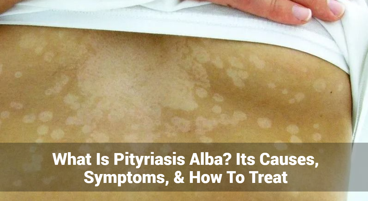
Pityriasis alba is a skin disorder that manifests as pale or light-colored patches on the skin. These patches are usually slightly scaly and have well-defined borders. The condition is most commonly observed in children and adolescents but can also affect adults. Although it often resolves on its own over time, the condition can cause cosmetic concerns and anxiety.
What Causes of Pityriasis Alba?
The exact cause of pityriasis alba is not fully understood, but several factors are believed to contribute to its development:
- Skin Dryness: One of the most common theories is that pityriasis alba is related to skin dryness. The patches may appear as a result of the skin’s reduced ability to retain moisture, leading to a contrast between the affected and unaffected areas.
- Sun Exposure: There is evidence to suggest that increased sun exposure can exacerbate or trigger pityriasis alba. The skin patches often become more noticeable after sun exposure, which can further lighten the affected areas.
- Eczema: Pityriasis alba may be associated with a history of eczema (atopic dermatitis). Children with eczema may develop these patches as a residual effect of their eczema.
- Immunological Factors: Some researchers believe that pityriasis alba may be related to an immune response that affects skin pigmentation, although more research is needed to confirm this theory.
- Nutritional Deficiencies: While not a primary cause, certain nutritional deficiencies, particularly in vitamins like vitamin D and calcium, may influence the appearance of pityriasis alba.
Track and Manage your Eczema treatment using a comprehensive Eczema App
Download Eczemaless now
![]()
![]()
![]()
![]()
![]()
![]()
![]()
![]()
![]()
![]()
![]()
![]()
![]()
![]()
![]()
![]()
![]()
![]()
![]()
![]()
![]()
![]()
![]()
What Are The Common Symptoms of Pityriasis Alba?
The symptoms of pityriasis alba are typically mild and may include:
- Light-Colored Patches: The primary symptom is the presence of light-colored, slightly scaly patches on the skin. These patches are usually paler than the surrounding skin and have well-defined borders.
- Dry, Flaky Skin: The affected skin may appear dry and flaky, although this is not always the case. The patches may be more noticeable in individuals with darker skin tones.
- Asymptomatic: In most cases, pityriasis alba is asymptomatic, meaning it does not cause itching, pain, or other discomfort. However, some individuals may experience mild itching or irritation.
- Common Locations: Pityriasis alba commonly appears on the face, arms, and torso. The patches may be more visible in areas that are frequently exposed to the sun.
Diagnosis of Pityriasis Alba
Diagnosing pityriasis alba typically involves a physical examination by a dermatologist. The doctor will examine the skin patches and assess their characteristics. In some cases, a skin biopsy may be performed to rule out other skin conditions that may present with similar symptoms, such as vitiligo or tinea versicolor.
Treatment Options for Pityriasis Alba
Treatment for pityriasis alba is generally not necessary, as the condition often resolves on its own over time. However, there are several treatment options available to improve the appearance of the skin and manage symptoms:
1. Moisturizers: Regular application of moisturizers can help alleviate dryness and improve the appearance of affected skin. Products containing emollients and humectants, such as ceramides or hyaluronic acid, are particularly effective in maintaining skin hydration.
2. Topical Steroids: In cases where there is significant dryness or irritation, topical corticosteroids may be prescribed. These medications help reduce inflammation and improve the appearance of the skin. Low-potency corticosteroids are typically used for this purpose.
3. Sunscreen: Using sunscreen is crucial for managing pityriasis alba. Sunscreen helps protect the skin from further sun damage and prevents the patches from becoming more noticeable. Broad-spectrum sunscreens with an SPF of 30 or higher are recommended.
4. Topical Calcineurin Inhibitors: For individuals with persistent symptoms, topical calcineurin inhibitors such as tacrolimus or pimecrolimus may be used. These medications help reduce inflammation without the side effects associated with steroids.
5. Pigment Restoring Treatments: Although not always necessary, some people seek treatments to restore skin pigmentation. Options include topical treatments containing ingredients like hydroquinone or laser therapy. These treatments should be discussed with a dermatologist to determine their suitability.
GET IN CONTROL OF YOUR ECZEMA
Use our AI tool to check the severity of Eczema and keep track of your Eczema progress.

Natural Remedies for Pityriasis Alba
In addition to conventional treatments, several natural remedies may help improve the appearance of pityriasis alba and support skin health:
1. Aloe Vera: Aloe vera has moisturizing and soothing properties that can benefit dry, flaky skin. Applying pure aloe vera gel to the affected areas may help improve hydration and reduce flakiness.
2. Coconut Oil: Coconut oil is known for its moisturizing and anti-inflammatory properties. Applying coconut oil to the skin can help alleviate dryness and improve skin texture.
3. Vitamin E: Vitamin E is a powerful antioxidant that supports skin health. Applying vitamin E oil or using skincare products containing vitamin E may help improve the appearance of the skin and protect it from damage.
4. Oatmeal Baths: Oatmeal has soothing properties that can help relieve dry and itchy skin. Taking an oatmeal bath or using colloidal oatmeal in a bath can provide relief and improve skin condition.
5. Proper Hydration: Drinking plenty of water and maintaining overall hydration is essential for skin health. Proper hydration helps keep the skin moisturized from within and may contribute to the overall improvement of skin appearance.
Lifestyle Tips for Managing Pityriasis Alba
In addition to treatments and remedies, adopting certain lifestyle changes can help manage and prevent pityriasis alba:
- Maintain Skin Hydration: Regularly apply moisturizers to keep the skin hydrated and prevent dryness.
- Protect from Sun Exposure: Use sunscreen and protective clothing to shield the skin from harmful UV rays, which can exacerbate the condition.
- Avoid Irritants: Be mindful of skincare products that may irritate the skin. Opt for gentle, non-irritating products suitable for sensitive skin.
- Healthy Diet: Maintain a balanced diet rich in vitamins and minerals to support overall skin health. Foods high in antioxidants and essential fatty acids can be beneficial.
- Regular Skin Care: Follow a consistent skincare routine that includes cleansing and moisturizing to keep the skin healthy and resilient.
Prognosis and Outlook
Pityriasis alba is a benign condition with a generally favorable prognosis. The light-colored patches often resolve on their own over time, although the process may take several months to years. In many cases, the skin returns to its normal pigmentation without the need for medical intervention.
Conclusion
Pityriasis alba is a common skin condition characterized by light-colored patches, often affecting children and adolescents. While it is benign and typically resolves on its own, understanding its causes, symptoms, and treatment options can help manage the condition more effectively. Regular moisturization, sun protection, and appropriate treatments can improve skin appearance and alleviate any associated discomfort. If you or someone you know is experiencing symptoms of pityriasis alba, consult a dermatologist OR healthcare professional can provide personalized advice and treatment options to ensure optimal skin health and management.
Track and Manage your Eczema treatment using a comprehensive Eczema App
Download Eczemaless now
![]()
![]()
![]()
![]()
![]()
![]()
![]()
![]()
![]()
![]()
![]()
![]()
![]()
![]()
![]()
![]()
![]()
![]()
![]()
![]()
![]()
![]()
![]()