How to Identify and Prevent Eczema Flare-Ups in Adults

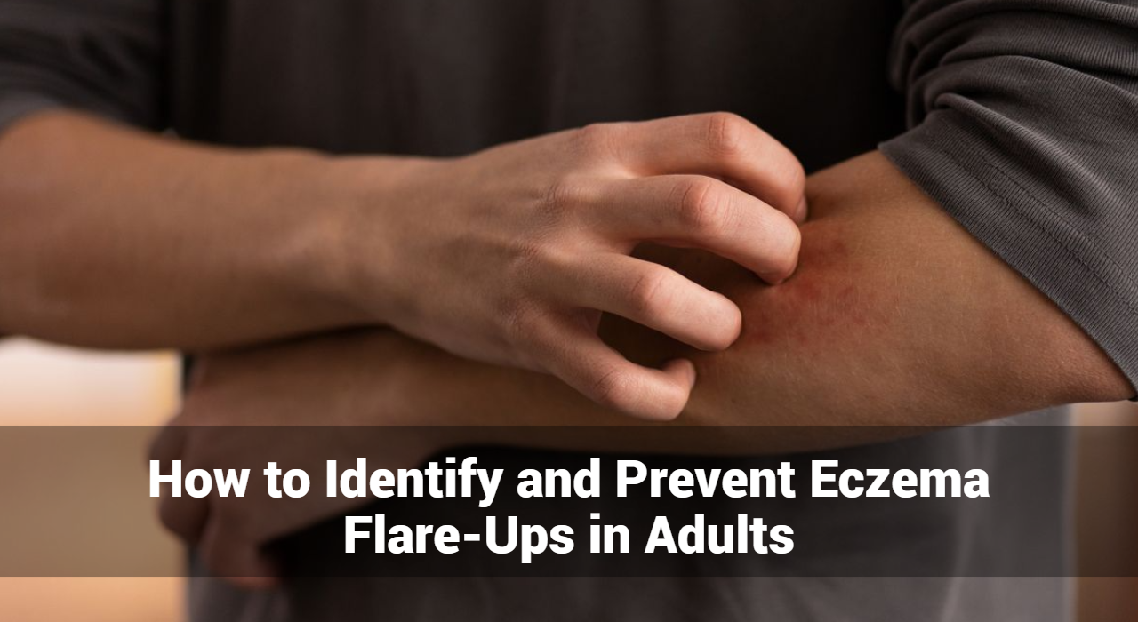
Eczema flare-ups can be a frustrating and uncomfortable experience for adults. Understanding how to identify and prevent these flare-ups is essential for managing the condition effectively. In this article, we will explore the common triggers, symptoms, and preventive measures for eczema flare-ups.

What is Eczema?

Eczema, also known as atopic dermatitis, is a condition that makes the skin red, inflamed, and itchy. It is a common condition that can occur at any age but is more frequently seen in children. Eczema can vary in severity and is often chronic, meaning it can last for a long time, with periods of remission and exacerbation.

What Are Eczema Flare-Ups?

Eczema flare-ups refer to periods when the symptoms of eczema, a chronic skin condition, become significantly worse. These flare-ups are characterized by increased inflammation, itching, redness, and other symptoms that can cause considerable discomfort. Understanding what triggers these flare-ups and how to manage them is essential for individuals living with eczema.

Identifying Eczema Flare-Ups

Eczema flare-ups can be a distressing experience, characterized by a sudden worsening of symptoms. Understanding how to identify these flare-ups is crucial for effective management and timely intervention. In this article, we will delve into the signs, triggers, and strategies for recognizing eczema flare-ups.

Recognizing the Signs of Eczema Flare-Ups

Eczema flare-ups present with a variety of symptoms, often varying in intensity. Here are the key signs to look out for:

  1. Red, Inflamed Skin
    • One of the most noticeable signs of an eczema flare-up is red, swollen patches of skin. These areas may feel warm to the touch and appear puffy.
  2. Intense Itching
    • Itching is a hallmark symptom of eczema. During a flare-up, the itching can become severe, leading to constant scratching that worsens the condition.
  3. Dry, Scaly Patches
    • Flare-ups often cause the skin to become extremely dry and scaly. These patches can be rough and may flake off.
  4. Blisters and Oozing
    • In some cases, small, fluid-filled blisters may form. These blisters can burst, leading to oozing and crusting over.
  5. Cracked and Painful Skin
    • Severely dry skin can crack, creating painful fissures that are susceptible to infection. This can lead to further complications if not treated promptly.
  6. Thickened Skin
    • Chronic scratching and rubbing can cause the skin to thicken and develop a leathery texture, a condition known as lichenification.

Track and Manage your Eczema treatment using a comprehensive Eczema App
Download Eczemaless now


What Are Common Causes of Eczema Flare-Ups in Adults?

Several factors can trigger eczema flare-ups in adults, including:

  1. Environmental Irritants
    • Chemicals: Exposure to harsh chemicals in cleaning products, soaps, and detergents can irritate the skin.
    • Pollution: Airborne pollutants can exacerbate eczema symptoms.
  2. Allergens
    • Pollen and Dust: Common allergens like pollen and dust mites can trigger eczema flare-ups.
    • Pet Dander: Proteins found in pet dander can cause allergic reactions leading to eczema.
  3. Weather Conditions
    • Cold Weather: Dry, cold air can strip the skin of moisture, leading to flare-ups.
    • Hot Weather: Heat and sweat can irritate the skin and cause itching.
  4. Stress
    • Emotional stress can worsen eczema symptoms by triggering the body’s inflammatory response.
  5. Diet
    • Certain foods, such as dairy, nuts, and wheat, can trigger eczema in some individuals.
  6. Hormonal Changes
    • Fluctuations in hormone levels, particularly in women, can lead to eczema flare-ups.

How Can Prevent Eczema Flare-Ups For Adults?

Preventing eczema flare-ups involves a combination of lifestyle adjustments and skincare routines:

  1. Moisturize Regularly
    • Use fragrance-free moisturizers to keep the skin hydrated and prevent dryness. Apply immediately after bathing to lock in moisture.
  2. Avoid Triggers
    • Identify and avoid known triggers, such as specific foods, allergens, and irritants. Keeping a diary can help track what causes flare-ups.
  3. Manage Stress
    • Practice stress-reducing techniques such as yoga, meditation, and deep breathing exercises.
  4. Wear Protective Clothing
    • Use gloves when handling chemicals or cleaning products. Wear loose, breathable clothing to reduce skin irritation.
  5. Use Gentle Skin Care Products
    • Opt for hypoallergenic, fragrance-free soaps, and detergents. Avoid products with harsh chemicals that can irritate the skin.
  6. Maintain a Healthy Diet
    • Eat a balanced diet rich in fruits, vegetables, and omega-3 fatty acids to support skin health.
  7. Stay Hydrated
    • Drink plenty of water to keep your skin hydrated from the inside out.
  8. Monitor Weather Conditions
    • Protect your skin in extreme weather conditions by using a humidifier in dry, cold climates and staying cool in hot weather.


GET IN CONTROL OF YOUR ECZEMA

Use our AI tool to check the severity of Eczema and keep track of your Eczema progress.

Use our AI tool to check the severity of Eczema and keep track of your Eczema progress.


Home Remedies for Eczema Flare-Ups in Adults

Managing eczema flare-ups at home can significantly reduce discomfort and improve skin health. While medical treatments are often necessary for severe cases, many individuals find relief through simple, natural remedies. Here are some effective home remedies for managing eczema flare-ups in adults.

1. Moisturize Regularly

Hydration is key to managing eczema. Keeping the skin moisturized helps to prevent dryness and flakiness.

  • Use Thick Moisturizers: Opt for thick creams or ointments rather than lotions. Look for products containing ingredients like ceramides, glycerin, or hyaluronic acid.
  • Apply After Bathing: Apply moisturizer immediately after bathing to lock in moisture.

2. Warm Baths

Soothing baths can help alleviate itching and inflammation.

  • Oatmeal Baths: Adding colloidal oatmeal to a lukewarm bath can soothe the skin and reduce itching. Oatmeal has anti-inflammatory properties that can provide relief.
  • Epsom Salt Baths: Epsom salt or Dead Sea salt baths can help reduce inflammation and promote healing.

3. Coconut Oil

Coconut oil has antimicrobial and anti-inflammatory properties.

  • How to Use: Apply virgin coconut oil directly to the affected areas to help moisturize and soothe the skin.

4. Aloe Vera

Aloe vera gel is known for its soothing and healing properties.

  • How to Use: Apply pure aloe vera gel from the plant or use a store-bought gel with minimal additives. This can help reduce redness and itching.

5. Honey

Honey is a natural antibacterial and anti-inflammatory agent.

  • How to Use: Apply raw honey to the affected areas and leave it on for 20-30 minutes before rinsing off. This can help promote healing and reduce flare-ups.

6. Apple Cider Vinegar

Apple cider vinegar can help restore the skin’s natural acidity and fight bacteria.

  • How to Use: Dilute apple cider vinegar with equal parts water and apply to the skin using a cotton ball. Rinse off after 5-10 minutes. Be cautious, as it may sting on open or cracked skin.

7. Tea Tree Oil

Tea tree oil has anti-inflammatory and antiseptic properties.

  • How to Use: Dilute a few drops of tea tree oil with a carrier oil (like coconut or olive oil) and apply to the affected areas. Avoid using undiluted tea tree oil directly on the skin, as it can cause irritation.

8. Dietary Adjustments

Healthy diet changes can have a positive impact on eczema management.

  • Anti-Inflammatory Foods: Include foods rich in omega-3 fatty acids (like fish, flaxseeds, and walnuts), antioxidants (like berries, leafy greens, and nuts), and probiotics (like yogurt and fermented foods).
  • Avoid Triggers: Identify and avoid foods that trigger flare-ups. Common triggers include dairy, gluten, and processed foods.

9. Wet Wrap Therapy

Wet wraps can help hydrate and soothe severely dry skin.

  • How to Use: After applying moisturizer or medication, cover the affected area with a damp cloth or bandage. Then, cover with a dry layer. Leave on for several hours or overnight.

10. Manage Stress

Stress management techniques can help reduce the frequency and severity of flare-ups.

  • Relaxation Techniques: Practice yoga, meditation, deep breathing exercises, or other relaxation techniques to manage stress.

11. Maintain Proper Hygiene

Good hygiene practices can prevent infections and reduce flare-ups.

  • Mild Cleansers: Use mild, fragrance-free cleansers that don’t strip the skin of its natural oils.
  • Avoid Hot Water: Use lukewarm water for bathing and avoid hot showers, which can dry out the skin.

12. Clothing Choices

Choosing the right clothing can help minimize skin irritation.

  • Soft Fabrics: Wear loose-fitting clothes made from soft fabrics like cotton. Avoid scratchy materials like wool.
  • Breathable Fabrics: Choose breathable fabrics to reduce sweating and irritation.

Conclusion

By understanding the common triggers and symptoms of eczema flare-ups in adults can take proactive steps to manage their condition and prevent flare-ups. Regular moisturizing, avoiding known triggers, managing stress, and using gentle skincare products are key strategies in maintaining healthy skin. If you continue to experience severe flare-ups, consult a dermatologist for personalized treatment options.


Track and Manage your Eczema treatment using a comprehensive Eczema App
Download Eczemaless now


Occupational Dermatitis: Types, Causes, Symptoms & Treatments

Occupational dermatitis is a common work-related skin condition resulting from exposure to irritants or allergens in the workplace. It significantly impacts the quality of life and productivity of affected individuals. Understanding the types, causes, symptoms, and treatments is essential for effective management and prevention.

What is Occupational Dermatitis?

Occupational dermatitis is a type of skin inflammation caused or aggravated by workplace exposures. It is one of the most common occupational diseases and can significantly impact an individual’s quality of life and work productivity.

What Are The Types of Occupational Dermatitis?

Occupational dermatitis is a broad term for skin conditions caused or aggravated by workplace exposures. There are two main types of occupational dermatitis: irritant contact dermatitis and allergic contact dermatitis. Here’s an overview of each type, including their causes, symptoms, and treatments.

1. Irritant Contact Dermatitis (ICD)

Irritant Contact Dermatitis (ICD) is a type of skin inflammation caused by direct damage to the skin from contact with irritants. It is the most common form of occupational dermatitis and can occur in any work environment where the skin is exposed to harsh substances or conditions.

Causes:

ICD results from direct damage to the skin by chemical, physical, or biological agents. Common causes include:

  • Chemical Irritants: Acids, alkalis, solvents, detergents, and disinfectants. Frequent exposure to these substances can disrupt the skin barrier, leading to inflammation and irritation.
  • Physical Irritants: Friction, pressure, extreme temperatures (heat and cold), and prolonged exposure to water (wet work). These factors can cause mechanical damage to the skin.
  • Biological Irritants: Bodily fluids such as blood and saliva, particularly in healthcare settings, can cause dermatitis.

Symptoms:

  • Redness and Swelling: The affected area often becomes red and swollen.
  • Dry, Cracked Skin: Skin may appear dry, cracked, and scaly.
  • Pain and Itching: The area may be painful or itchy.
  • Blisters: In severe cases, blisters can form.

Treatment:

  • Avoidance of Irritants: Identifying and avoiding contact with the irritant.
  • Barrier Creams and Moisturizers: Regular application to protect and repair the skin.
  • Topical Steroids: Used to reduce inflammation and promote healing.
  • Proper Hand Hygiene: Using gentle cleansers and moisturizing frequently.

2. Allergic Contact Dermatitis (ACD)

Allergic Contact Dermatitis (ACD) is an inflammatory skin condition caused by an immune reaction to a substance (allergen) that comes into contact with the skin. This type of occupational dermatitis involves a delayed hypersensitivity reaction, typically occurring 24 to 72 hours after exposure.

Causes:

ACD occurs when the skin becomes sensitized to an allergen, leading to an immune response upon subsequent exposures. Common causes include:

  • Metals: Nickel, cobalt, and chromium, often found in tools, machinery, and jewelry.
  • Latex: Used in gloves and other medical supplies, common in healthcare and laboratory environments.
  • Fragrances and Preservatives: Found in personal care products, cosmetics, and industrial products.
  • Rubber Additives: Chemicals used in the manufacture of rubber products like gloves and handles.
  • Dyes and Resins: Used in textiles, printing, and manufacturing industries.
  • Plants: Certain plants and plant products, such as poison ivy, can cause allergic reactions.

Symptoms:

  • Rash and Redness: The affected area develops a red rash.
  • Blisters and Oozing: Blisters may form and ooze fluid.
  • Swelling: The area may become swollen and inflamed.
  • Itching and Burning: Severe itching and a burning sensation are common.

Treatment:

  • Identification and Avoidance: Identifying the allergen through patch testing and avoiding exposure.
  • Topical Corticosteroids: Applied to reduce inflammation and itching.
  • Oral Antihistamines: Used to relieve itching.
  • Emollients: Moisturizers to soothe and repair the skin barrier.

Other Types of Occupational Dermatitis


Track and Manage your Eczema treatment using a comprehensive Eczema App
Download Eczemaless now


3. Photoallergic Contact Dermatitis

Photoallergic Contact Dermatitis is a type of skin inflammation triggered by a reaction between a chemical on the skin and exposure to ultraviolet (UV) light. This condition involves a delayed hypersensitivity reaction, usually manifesting 24 to 72 hours after exposure to both the allergen and sunlight.

Causes of Photoallergic Contact Dermatitis:

  1. Topical Medications:
    • Sunscreens: Certain chemical ingredients like oxybenzone.
    • Antibiotics: Topical antibiotics such as sulfonamides.
    • NSAIDs: Nonsteroidal anti-inflammatory drugs applied to the skin.
  2. Personal Care Products:
    • Fragrances: Found in perfumes and lotions.
    • Preservatives: Chemicals used to extend the shelf life of cosmetics and skin care products.
  3. Industrial Chemicals:
    • Coal Tar: Used in treatments for skin conditions like psoriasis.
    • Dyes: Chemicals used in various industrial processes.

Symptoms of Photoallergic Contact Dermatitis:

  • Redness and Swelling: Inflammation typically in areas exposed to sunlight.
  • Itching and Burning: The affected skin can be intensely itchy and may have a burning sensation.
  • Blisters: Fluid-filled blisters that may ooze and crust over.
  • Eczema-like Rash: The skin may develop a rash that resembles eczema.

Treatment of Photoallergic Contact Dermatitis:

  • Avoidance of Allergen and Sunlight: Identifying and avoiding the specific chemical causing the reaction, and limiting exposure to sunlight.
  • Topical Corticosteroids: Reducing inflammation and relieving itching.
  • Oral Antihistamines: Used to manage itching and discomfort.
  • Cool Compresses: Soothe the skin and reduce inflammation.
  • Emollients: Help to repair the skin barrier and prevent dryness.

4. Contact Urticaria

Contact urticaria, also known as hives, is an immediate allergic reaction that occurs when the skin comes into direct contact with an allergen or irritant. Unlike other forms of contact dermatitis that may take hours or days to develop, contact urticaria typically appears within minutes of exposure.

Causes of Contact Urticaria

Contact urticaria can be triggered by a wide variety of substances, which can be classified into two categories: immunologic and non-immunologic.

Immunologic (Allergic) Contact Urticaria:

  • Proteins: Latex, animal dander, and certain foods (e.g., fruits, vegetables, and seafood).
  • Plants: Nettle, some herbs, and spices.
  • Chemicals: Preservatives, fragrances, and certain medications applied to the skin.

Non-Immunologic (Irritant) Contact Urticaria:

  • Chemicals: Organic solvents, certain industrial chemicals, and some cosmetics.
  • Physical Agents: Cold, heat, and pressure can also provoke a non-immunologic reaction.

Symptoms of Contact Urticaria

Symptoms of contact urticaria are typically localized to the area of contact but can spread, especially in severe cases. They include:

  • Hives (Urticaria): Red, raised, itchy welts that can vary in size and shape.
  • Itching and Burning: Intense itching and a burning sensation at the site of contact.
  • Swelling (Angioedema): Swelling of deeper layers of the skin, particularly around the eyes, lips, and throat.
  • Systemic Symptoms: In severe cases, symptoms can include anaphylaxis, characterized by difficulty breathing, rapid heartbeat, and a drop in blood pressure, requiring immediate medical attention.

Treatment of Contact Urticaria

The primary treatment goals are to relieve symptoms and prevent future reactions:

  • Avoidance of Triggers: Identifying and avoiding contact with known allergens and irritants.
  • Antihistamines: Used to relieve itching and reduce hives.
  • Corticosteroids: Topical or oral corticosteroids to reduce inflammation.
  • Epinephrine: For severe reactions or anaphylaxis, an epinephrine auto-injector (e.g., EpiPen) is essential.
  • Cool Compresses: Applying cool compresses to the affected area can provide symptomatic relief.

What Are The Common Workplace Sources and Risk Factors?

  • Healthcare: Frequent handwashing, use of latex gloves, and exposure to disinfectants.
  • Construction and Manufacturing: Contact with cement, solvents, and other industrial chemicals.
  • Hairdressing and Beauty: Exposure to dyes, bleaches, and other hair and skin products.
  • Food Industry: Prolonged exposure to water, detergents, and food products.
  • Agriculture: Contact with pesticides, fertilizers, and plant allergens.


GET IN CONTROL OF YOUR ECZEMA

Use our AI tool to check the severity of Eczema and keep track of your Eczema progress.

Use our AI tool to check the severity of Eczema and keep track of your Eczema progress.


Preventive Measures From Occupational Dermatitis

  • Personal Protective Equipment (PPE): Gloves, protective clothing, and barrier creams.
  • Workplace Controls: Implementing engineering controls to minimize exposure, such as ventilation systems and safe handling procedures.
  • Education and Training: Educating workers about risks, proper skin care, and safe handling practices.
  • Skin Care: Regular use of moisturizers and mild skin cleansers to maintain skin barrier integrity.

While professional medical advice and proper workplace safety measures are crucial for managing and preventing occupational dermatitis, natural remedies can also help alleviate symptoms and promote skin healing. Here are some natural remedies that might be beneficial:

Natural Remedies for Occupational Dermatitis

While professional medical advice and proper workplace safety measures are crucial for managing and preventing occupational dermatitis, natural remedies can also help alleviate symptoms and promote skin healing. Here are some natural remedies that might be beneficial:

  1. Aloe Vera
    • Benefits: Aloe vera has anti-inflammatory and soothing properties.
    • Usage: Apply pure aloe vera gel directly to the affected areas. Ensure it’s 100% aloe vera without added fragrances or alcohol.
  2. Coconut Oil
    • Benefits: Coconut oil is a natural moisturizer with antimicrobial properties.
    • Usage: Apply virgin coconut oil to the skin to keep it hydrated and help repair the skin barrier.
  3. Oatmeal Baths
    • Benefits: Colloidal oatmeal can relieve itching and inflammation.
    • Usage: Add finely ground oatmeal to a lukewarm bath and soak for 15-20 minutes.
  4. Chamomile
    • Benefits: Chamomile has anti-inflammatory and soothing effects.
    • Usage: Apply chamomile tea bags or a compress soaked in chamomile tea to the affected areas.
  5. Honey
    • Benefits: Honey has antibacterial and wound-healing properties.
    • Usage: Apply raw honey to the affected skin, leave it on for 15-20 minutes, and then rinse off gently with lukewarm water.
  6. Calendula
    • Benefits: Calendula has anti-inflammatory and healing properties.
    • Usage: Use calendula ointment or cream on the affected areas.
  7. Tea Tree Oil
    • Benefits: Tea tree oil has anti-inflammatory and antimicrobial properties.
    • Usage: Dilute tea tree oil with a carrier oil (like coconut oil) before applying to the skin. Do a patch test first to check for any sensitivity.
  8. Cucumber Slices
    • Benefits: Cucumbers are soothing and can reduce inflammation.
    • Usage: Place slices of fresh cucumber on the affected areas for a cooling effect.
  9. Apple Cider Vinegar
    • Benefits: Apple cider vinegar has antibacterial and antifungal properties.
    • Usage: Dilute apple cider vinegar with water (1 part vinegar to 3 parts water) and apply with a cotton ball. Avoid using it on open or cracked skin.
  10. Olive Oil
    • Benefits: Olive oil is a natural moisturizer rich in antioxidants.
    • Usage: Apply extra virgin olive oil to the skin to help retain moisture and promote healing.

Additional Tips

  • Stay Hydrated: Drinking plenty of water helps maintain overall skin health.
  • Diet: A diet rich in antioxidants, vitamins, and omega-3 fatty acids can support skin healing. Include foods like fruits, vegetables, nuts, and fatty fish.
  • Avoid Triggers: Identify and avoid substances that trigger your dermatitis.

Conclusion

Managing occupational dermatitis involves a combination of preventive measures, early detection, and appropriate treatment. Collaboration between employers and employees is crucial to create a safe and healthy work environment. Always consult with a healthcare provider for personalized diagnosis and treatment plans.


Track and Manage your Eczema treatment using a comprehensive Eczema App
Download Eczemaless now


What Is Dyshidrotic Eczema? Its Symptoms, Causes, Treatment, & Prevention

Dyshidrotic eczema, also known as pompholyx or dyshidrosis, is a type of eczema that affects the hands and feet. Characterized by small, itchy blisters, dyshidrotic eczema can be a chronic and recurring condition that significantly impacts a person’s quality of life. This article provides a comprehensive overview of dyshidrotic eczema, including its symptoms, causes, treatment options, and preventive measures.

What Is Dyshidrotic Eczema?

Dyshidrotic eczema is a skin condition that primarily affects the palms of the hands, sides of the fingers, and soles of the feet. The condition manifests as small, fluid-filled blisters that cause intense itching and discomfort. These blisters can persist for several weeks, causing significant pain and potentially leading to skin cracking and infection.

Epidemiology

Dyshidrotic eczema is relatively common, affecting approximately 1 in 5,000 people. It can occur at any age but is most frequently seen in adults aged 20 to 40. The condition tends to be more prevalent in warmer climates and during spring and summer months.

What Are The Different Types of Dyshidrotic Eczema?

Dyshidrotic eczema, also known as pompholyx, is a specific type of eczema that affects the hands and feet. While it is generally considered a single condition, dyshidrotic eczema can present in various forms, which can be classified based on severity, chronicity, and underlying triggers. Here, we’ll explore these variations in detail.

1. Acute Dyshidrotic Eczema

Characteristics:

  • Sudden onset of symptoms.
  • Development of small, intensely itchy blisters on the palms, sides of the fingers, and soles of the feet.
  • The blisters are often filled with a clear fluid and can cause significant discomfort.

Causes:

  • Acute flare-ups may be triggered by stress, allergens, or exposure to irritants.
  • Changes in weather or humidity levels can also provoke an acute episode.

Symptoms:

  • Intense itching and burning sensation.
  • Redness and swelling around the blistered areas.

Treatment:

  • Topical corticosteroids to reduce inflammation.
  • Cold compresses to alleviate itching.
  • Avoiding known triggers to prevent future flare-ups.

2. Chronic Dyshidrotic Eczema

Characteristics:

  • Persistent or recurring episodes over a long period.
  • Skin thickening (lichenification) and fissures due to chronic scratching and inflammation.

Causes:

  • Ongoing exposure to irritants or allergens.
  • Underlying medical conditions like atopic dermatitis.

Symptoms:

  • Continuous or frequently recurring blisters.
  • Dry, cracked, and thickened skin.

Treatment:

  • Long-term use of emollients to maintain skin hydration.
  • Topical or oral corticosteroids during flare-ups.
  • Lifestyle modifications to minimize exposure to triggers.


Track and Manage your Eczema treatment using a comprehensive Eczema App
Download Eczemaless now


3. Recurrent Dyshidrotic Eczema

Characteristics:

  • Episodes that occur in cycles, with periods of remission in between.
  • Blisters reappear periodically, often without an apparent trigger.

Causes:

  • Allergens, stress, or other environmental factors that periodically affect the patient.

Symptoms:

  • Similar to acute dyshidrotic eczema but with intermittent flare-ups.
  • Clear fluid-filled blisters, itching, and redness.

Treatment:

  • Preventive measures during remission phases, such as maintaining a consistent skincare routine.
  • Quick intervention with topical treatments at the onset of symptoms.

4. Hyperkeratotic Dyshidrotic Eczema

Characteristics:

  • Development of thick, scaly patches on the hands and feet.
  • Often accompanied by fewer blisters compared to other forms.

Causes:

  • Chronic irritation and inflammation.
  • Genetic predisposition to hyperkeratosis.

Symptoms:

  • Thickened, scaly skin that can crack and bleed.
  • Mild to moderate itching.

Treatment:

  • Keratolytic agents like salicylic acid to reduce skin thickening.
  • Emollients to soften the skin.
  • Topical steroids for inflammation.

5. Vesicular Dyshidrotic Eczema

Characteristics:

  • Presence of numerous small vesicles (blisters) filled with clear fluid.
  • Blisters are more numerous and can merge to form larger bullae.

Causes:

  • Similar triggers as other forms, including allergens, stress, and irritants.

Symptoms:

  • Intense itching and burning sensation.
  • Blisters that may rupture, leading to crusting and potential secondary infection.

Treatment:

  • Antihistamines to control itching.
  • Antiseptic soaks to prevent infection.
  • Topical steroids for severe inflammation.

6. Infective Dyshidrotic Eczema

Characteristics:

  • Secondary bacterial or fungal infection superimposed on dyshidrotic eczema.
  • Increased severity and risk of complications.

Causes:

  • Open blisters and cracked skin can become infected with bacteria (e.g., Staphylococcus aureus) or fungi (e.g., Candida species).

Symptoms:

  • Increased redness, swelling, and pain.
  • Pus formation and potential fever.

Treatment:

  • Antibiotics or antifungal medications to treat the infection.
  • Continued use of moisturizers and topical steroids once the infection is controlled.

7. Allergic Dyshidrotic Eczema

Characteristics:

  • Triggered by an allergic reaction to substances such as metals (nickel), certain foods, or topical products.
  • Blisters appear after contact with or ingestion of the allergen.

Causes:

  • Allergic contact dermatitis can provoke dyshidrotic eczema in susceptible individuals.

Symptoms:

  • Blistering, itching, and redness localized to areas in contact with the allergen.
  • Systemic symptoms if caused by ingested allergens.

Treatment:

  • Identification and avoidance of the allergen.
  • Topical steroids to reduce allergic inflammation.
  • Antihistamines to control itching.

Symptoms of Dyshidrotic Eczema

Primary Symptoms

  1. Blisters: Small, fluid-filled blisters that typically appear on the fingers, palms, and soles. These blisters can be extremely itchy and painful.
  2. Itching: Intense itching is a hallmark of dyshidrotic eczema, often preceding the appearance of blisters.
  3. Redness: The affected areas may become red and inflamed.
  4. Cracking and Peeling: As blisters heal, the skin may crack, peel, and become painful.

Secondary Symptoms

  1. Pain: Severe itching and blistering can lead to significant pain, especially if the skin cracks or becomes infected.
  2. Swelling: Affected areas may swell due to inflammation and fluid buildup.
  3. Dryness: Skin may become dry and scaly after blisters resolve.
  4. Infection: Open blisters and cracked skin can become infected, leading to increased pain, pus formation, and other complications.

Complications

  1. Skin Infections: Bacterial infections can develop if blisters or cracked skin are not properly managed.
  2. Scarring: Repeated episodes and chronic inflammation can lead to permanent skin changes and scarring.
  3. Psychosocial Impact: Persistent symptoms can affect mental health, causing stress, anxiety, and social embarrassment.


GET IN CONTROL OF YOUR ECZEMA

Use our AI tool to check the severity of Eczema and keep track of your Eczema progress.

Use our AI tool to check the severity of Eczema and keep track of your Eczema progress.

What Are The Causes of Dyshidrotic Eczema?

Genetic Factors

  1. Family History: A family history of eczema or other atopic conditions can increase the risk of developing dyshidrotic eczema.
  2. Genetic Mutations: Certain genetic mutations, particularly those affecting skin barrier function, may predispose individuals to this condition.

Environmental Factors

  1. Allergens: Exposure to allergens such as pollen, dust mites, and pet dander can trigger dyshidrotic eczema in susceptible individuals.
  2. Irritants: Contact with irritants like soaps, detergents, and chemicals can exacerbate symptoms.
  3. Climate: Warm, humid climates and seasonal changes can influence the severity and frequency of outbreaks.

Lifestyle Factors

  1. Stress: Psychological stress is a known trigger for dyshidrotic eczema flare-ups.
  2. Diet: Certain foods, such as those high in nickel or cobalt, may trigger symptoms in some individuals.
  3. Hygiene Practices: Excessive washing and use of harsh skin products can damage the skin barrier and exacerbate symptoms.

Medical Conditions

  1. Atopic Dermatitis: Individuals with a history of atopic dermatitis are more likely to develop dyshidrotic eczema.
  2. Allergies: Allergic conditions, including hay fever and asthma, are often associated with dyshidrotic eczema.
  3. Infections: Fungal infections on the feet or hands can trigger dyshidrotic eczema in some cases.

Diagnosis of Dyshidrotic Eczema

Clinical Evaluation

  1. Medical History: A detailed medical history, including any family history of eczema or allergies, helps in diagnosing dyshidrotic eczema.
  2. Physical Examination: A dermatologist will examine the skin, noting the characteristic blisters and pattern of distribution.

Diagnostic Tests

  1. Skin Biopsy: A biopsy may be performed to rule out other conditions that mimic dyshidrotic eczema, such as contact dermatitis or fungal infections.
  2. Patch Testing: Patch testing can identify specific allergens that may be triggering the eczema.
  3. Blood Tests: Blood tests may be used to check for underlying conditions or infections that could be contributing to symptoms.

Treatment of Dyshidrotic Eczema

Topical Treatments

  1. Corticosteroids: Topical corticosteroids are commonly prescribed to reduce inflammation and itching. They are applied directly to the affected areas.
  2. Calcineurin Inhibitors: Non-steroidal options such as tacrolimus and pimecrolimus can help manage symptoms with fewer side effects than steroids.
  3. Moisturizers: Emollients and moisturizers are essential for maintaining skin hydration and barrier function.

Oral Medications

  1. Antihistamines: Oral antihistamines can help reduce itching and improve sleep.
  2. Corticosteroids: For severe cases, oral corticosteroids may be prescribed to quickly control inflammation.
  3. Immunosuppressants: Medications like cyclosporine or methotrexate may be used in severe, refractory cases.

Phototherapy

  1. UV Therapy: Ultraviolet light therapy, particularly narrowband UVB, can be effective in reducing symptoms for some patients.

Lifestyle and Home Remedies of Dyshidrotic Eczema

  1. Cold Compresses: Applying cold compresses to affected areas can reduce itching and inflammation.
  2. Avoiding Triggers: Identifying and avoiding triggers, such as certain foods, stress, or allergens, can help manage symptoms.
  3. Good Hygiene Practices: Using mild soaps and moisturizers, and avoiding excessive washing, can help protect the skin barrier.

Alternative Treatments

  1. Natural Remedies: Some patients find relief using natural treatments such as coconut oil, aloe vera, or apple cider vinegar.
  2. Acupuncture: Although not widely studied, some individuals report benefits from acupuncture.

Preventive Measures

  1. Skin Care Routine: Establishing a consistent skin care routine with gentle, hydrating products is crucial.
  2. Protective Clothing: Wearing gloves when handling irritants and breathable fabrics can help prevent flare-ups.
  3. Stress Management: Techniques such as yoga, meditation, and counseling can help manage stress levels.

Conclusion

Dyshidrotic eczema is a challenging condition that requires a multifaceted approach to management. Understanding the symptoms, causes, and treatment options can empower patients to take control of their condition and improve their quality of life. By working closely with a dermatologist and implementing preventive measures, individuals with dyshidrotic eczema can achieve better skin health and reduce the impact of this chronic condition on their daily lives.


Track and Manage your Eczema treatment using a comprehensive Eczema App
Download Eczemaless now


What Is Seborrheic Dermatitis? It’s Types, Cause, Symptoms, and Treatments

Seborrheic dermatitis is a common inflammatory skin condition that primarily affects areas of the body rich in oil glands, such as the scalp, face, and upper chest. It is characterized by redness, itching, and flaking of the skin, and it often occurs in areas with high sebum production. Let’s explore this condition in more detail:

What Is Seborrheic Dermatitis?

Seborrheic dermatitis is a common inflammatory skin condition that primarily affects areas of the body rich in oil glands, such as the scalp, face, and upper chest. It is characterized by redness, itching, and flaking of the skin, and it often occurs in areas with high sebum production. This condition can range from mild dandruff to more severe forms involving redness and scaling of the skin.

Seborrheic dermatitis is believed to be caused by a combination of factors, including the overgrowth of a yeast called Malassezia on the skin’s surface, excessive production of sebum (skin oil), genetic predisposition, hormonal changes, and environmental factors. While the exact cause is not fully understood, these factors are thought to contribute to the development of the condition.

Symptoms of seborrheic dermatitis can vary depending on the severity and location of the affected areas. Common symptoms include redness, scaling, itching, and flaking of the skin. On the scalp, seborrheic dermatitis often presents as dandruff-like flakes and itching, while on the face, it may appear as red patches with greasy scales, particularly in the eyebrows, nose, and ears.

Common Different Types of Seborrheic Dermatitis

Seborrheic dermatitis can manifest in various forms, affecting different areas of the body. While the underlying mechanism remains the same, the presentation may differ based on the location and severity of the condition. Here are the different types of seborrheic dermatitis:

  1. Scalp Seborrheic Dermatitis:
    • This type primarily affects the scalp, leading to dandruff-like flakes, redness, and itching.
    • Symptoms often include greasy or oily scales on the scalp, along with itching and irritation.
    • It can sometimes extend beyond the hairline onto the forehead or behind the ears.
  2. Facial Seborrheic Dermatitis:
    • Facial seborrheic dermatitis occurs on the face, particularly in areas rich in sebaceous glands, such as the eyebrows, sides of the nose, and around the mouth.
    • Symptoms include redness, greasy or scaly patches, and itching.
    • It may resemble other skin conditions like eczema or psoriasis, but it typically responds well to treatment.
  3. Trunk Seborrheic Dermatitis:
    • This type affects the trunk of the body, including the chest, back, and upper abdomen.
    • Symptoms include red, scaly patches on the skin, often with mild itching or discomfort.
    • Trunk seborrheic dermatitis may be less common than scalp or facial involvement but can still cause significant discomfort.
  4. Extremities Seborrheic Dermatitis:
    • Seborrheic dermatitis can also affect the extremities, including the arms and legs, although this is less common.
    • Symptoms may include redness, scaling, and mild itching on the affected areas.
    • Extremities seborrheic dermatitis may be associated with other forms of the condition or occur independently.
  5. Infantile Seborrheic Dermatitis (Cradle Cap):
    • This type affects infants, typically appearing within the first few months of life.
    • It presents as yellow, greasy scales on the scalp, often referred to as “cradle cap.”
    • Infantile seborrheic dermatitis is usually harmless and tends to resolve on its own within a few months without treatment.

While seborrheic dermatitis can affect various parts of the body, the underlying cause and treatment approach remain similar. Management typically involves the use of medicated shampoos, topical corticosteroids, antifungal agents, and moisturizers to control symptoms and prevent flare-ups. If you suspect you have seborrheic dermatitis, it’s important to consult a dermatologist for an accurate diagnosis and personalized treatment plan.


Track and Manage your Eczema treatment using a comprehensive Eczema App
Download Eczemaless now


Causes of Seborrheic Dermatitis:

Seborrheic dermatitis is a multifactorial condition with several potential causes contributing to its development. While the exact cause is not fully understood, several factors are believed to play a role in the onset and exacerbation of seborrheic dermatitis. Here are some of the primary causes and contributing factors:

  1. Malassezia Yeast Overgrowth:
    • Malassezia is a type of yeast that naturally resides on the skin’s surface. In individuals with seborrheic dermatitis, there is an overgrowth of Malassezia, which triggers an inflammatory response in the skin.
    • This yeast thrives in areas with high sebum (skin oil) production, such as the scalp, face, and upper chest, leading to the characteristic symptoms of seborrheic dermatitis.
  2. Excessive Sebum Production:
    • Seborrheic dermatitis often occurs in areas of the body with high sebum production, such as the scalp, face, and upper trunk.
    • Excessive sebum production can create an environment conducive to the growth of Malassezia yeast and contribute to the development of seborrheic dermatitis.
  3. Genetic Predisposition:
    • There appears to be a genetic component to seborrheic dermatitis, as it tends to run in families.
    • Certain genetic variations may predispose individuals to an exaggerated immune response to Malassezia yeast or alterations in skin barrier function, increasing the risk of developing seborrheic dermatitis.
  4. Hormonal Changes:
    • Hormonal fluctuations, such as those occurring during puberty, pregnancy, or menstrual cycles, may influence sebum production and contribute to the development or exacerbation of seborrheic dermatitis.
    • Hormonal imbalances can affect the skin’s oil glands and immune response, making individuals more susceptible to flare-ups of seborrheic dermatitis.
  5. Environmental Factors:
    • Environmental factors, such as cold weather, dry air, or humidity, can influence the severity of seborrheic dermatitis symptoms.
    • Exposure to certain environmental triggers, such as stress, fatigue, or certain skincare products, may exacerbate symptoms or trigger flare-ups in susceptible individuals.
  6. Immune System Dysfunction:
    • Dysfunction of the immune system may play a role in the pathogenesis of seborrheic dermatitis.
    • In individuals with seborrheic dermatitis, the immune system may overreact to the presence of Malassezia yeast, leading to inflammation and the characteristic symptoms of the condition.

While these factors are believed to contribute to the development of seborrheic dermatitis, the precise interplay between them and their relative importance may vary among individuals. Additionally, other factors, such as stress, diet, and medications, may also influence the severity and frequency of seborrheic dermatitis flare-ups.

What Are The Symptoms of Seborrheic Dermatitis?

Seborrheic dermatitis manifests through a variety of symptoms, which can range from mild to severe. The specific symptoms experienced by an individual may vary depending on the affected area of the body and the severity of the condition. Here are the common symptoms associated with seborrheic dermatitis:

  • Redness: Red patches or areas of erythema may develop on the skin, particularly in areas with inflammation or irritation.
  • Scaling: Flaky, white or yellowish scales may form on the skin’s surface, resembling dandruff. These scales may vary in size and can be easily shed from the affected areas.
  • Itching: Itching or pruritus is a common symptom of seborrheic dermatitis and can range from mild to severe. Itching may worsen in response to scratching and can contribute to further irritation of the skin.
  • Burning Sensation: Some individuals may experience a burning or stinging sensation on the skin, particularly in areas of active inflammation or irritation.
  • Greasy or Oily Skin: Affected areas of the skin may appear greasy or oily, especially on the scalp, face, or upper chest, where sebaceous glands are more abundant.
  • Erythema: Redness or inflammation of the skin may be present, particularly in areas where scaling and flaking occur.
  • Crusting: In severe cases, crusts or thick, adherent scales may form on the skin’s surface, especially on the scalp or in areas of chronic inflammation.
  • Hair Loss: In scalp seborrheic dermatitis, hair loss or thinning may occur, particularly if the condition is left untreated or becomes severe.
  • Sensitive Skin: The affected skin may become more sensitive or prone to irritation, making it uncomfortable to touch or manipulate.
  • Lesions: In some cases, seborrheic dermatitis may lead to the development of small, raised bumps or papules on the skin’s surface.

It’s important to note that the symptoms of seborrheic dermatitis can fluctuate over time, with periods of exacerbation and remission. Additionally, the severity of symptoms may vary among individuals and may be influenced by factors such as stress, environmental triggers, and underlying health conditions. If you experience persistent or severe symptoms of seborrheic dermatitis, it’s important to consult a dermatologist for an accurate diagnosis and appropriate treatment.


GET IN CONTROL OF YOUR ECZEMA

Use our AI tool to check the severity of Eczema and keep track of your Eczema progress.

Use our AI tool to check the severity of Eczema and keep track of your Eczema progress.

What is the most effective treatment for seborrheic dermatitis?

Treatment for seborrheic dermatitis aims to reduce inflammation, control symptoms, and prevent flare-ups. The choice of treatment depends on the severity of the condition and the areas of the body affected. Here are common treatment options for seborrheic dermatitis:

  1. Medicated Shampoos:
    • Over-the-counter shampoos containing active ingredients such as ketoconazole, selenium sulfide, coal tar, or zinc pyrithione can help reduce scaling, itching, and inflammation on the scalp.
    • These shampoos should be used regularly, typically two to three times per week, and left on the scalp for a few minutes before rinsing.
  2. Topical Corticosteroids:
    • Corticosteroid creams or lotions can be applied to affected areas of the skin to reduce inflammation and itching.
    • Low-potency corticosteroids are often used for facial seborrheic dermatitis, while higher-potency corticosteroids may be needed for more severe or resistant cases.
  3. Antifungal Creams or Lotions:
    • Topical antifungal medications, such as ketoconazole cream or ciclopirox olamine lotion, can help reduce the growth of Malassezia yeast on the skin and alleviate symptoms.
    • These medications are typically applied once or twice daily to affected areas of the skin.
  4. Calcineurin Inhibitors:
    • Topical calcineurin inhibitors, such as tacrolimus (Protopic) or pimecrolimus (Elidel), may be used as alternative treatments for seborrheic dermatitis, especially in sensitive areas like the face.
    • These medications work by suppressing inflammation and reducing immune system activity.
  5. Medicated Foams or Solutions:
    • Medicated foams or solutions containing corticosteroids, antifungal agents, or calcineurin inhibitors can be effective for treating seborrheic dermatitis on the scalp and other hairy areas.
    • These formulations are applied directly to the affected areas and can penetrate the hair follicles more effectively than creams or lotions.
  6. Oral Medications:
    • In severe or widespread cases of seborrheic dermatitis, oral medications such as oral antifungal agents (e.g., fluconazole) or systemic corticosteroids may be prescribed by a dermatologist.
    • Oral medications are typically reserved for cases that do not respond to topical treatments or for individuals with significant symptoms or complications.
  7. Moisturizers:
    • Regular use of moisturizers can help soothe and hydrate the skin, reducing flakiness and discomfort associated with seborrheic dermatitis.
    • Choose non-comedogenic moisturizers that won’t clog pores, especially for facial use.

It’s important to follow the recommendations of a healthcare professional when using these treatments, as some medications may have side effects or interactions with other medications. Additionally, long-term management may be necessary to control symptoms and prevent flare-ups of seborrheic dermatitis. If you have persistent or severe symptoms, consult a dermatologist for an accurate diagnosis and personalized treatment plan.

What Are The Natural Treatments (Home Remedies) For Seborrheic Dermatitis?

Natural treatments may help alleviate symptoms of seborrheic dermatitis and complement medical interventions. While they may not entirely replace conventional treatments, they can provide additional relief and promote overall skin health. Here are some natural remedies and self-care strategies for managing seborrheic dermatitis:

  • Regular Shampooing: Keeping the scalp clean is essential for managing seborrheic dermatitis. Use a mild, fragrance-free shampoo and wash your scalp regularly to remove excess oil and scales.
  • Tea Tree Oil: Tea tree oil has natural antifungal and anti-inflammatory properties that may help reduce symptoms of seborrheic dermatitis. Dilute tea tree oil with a carrier oil (such as coconut oil) and apply it to affected areas of the skin. Avoid using undiluted tea tree oil directly on the skin, as it can cause irritation.
  • Coconut Oil: Coconut oil has moisturizing and antimicrobial properties that may help soothe irritated skin and reduce inflammation. Apply virgin coconut oil to affected areas of the skin and scalp, leaving it on for a few hours or overnight before washing it off.
  • Apple Cider Vinegar: Apple cider vinegar has antimicrobial properties and can help restore the skin’s natural pH balance. Dilute apple cider vinegar with water and apply it to the scalp or affected areas of the skin as a rinse or compress.
  • Aloe Vera: Aloe vera gel has soothing and anti-inflammatory properties that can help alleviate itching and irritation associated with seborrheic dermatitis. Apply pure aloe vera gel to affected areas of the skin and leave it on for several minutes before rinsing it off.
  • Omega-3 Fatty Acids: Omega-3 fatty acids found in fatty fish, flaxseeds, and walnuts have anti-inflammatory properties that may help reduce symptoms of seborrheic dermatitis. Incorporate foods rich in omega-3 fatty acids into your diet or consider taking omega-3 supplements.
  • Probiotics: Probiotics are beneficial bacteria that can help support a healthy immune system and may reduce inflammation associated with seborrheic dermatitis. Consume probiotic-rich foods such as yogurt, kefir, sauerkraut, and kombucha, or take probiotic supplements.
  • Avoid Triggers: Identify and avoid triggers that may exacerbate symptoms of seborrheic dermatitis, such as stress, certain foods, alcohol, and harsh skincare products.
  • Manage Stress: Stress can worsen inflammation and trigger flare-ups of seborrheic dermatitis. Practice stress-reducing techniques such as yoga, meditation, deep breathing exercises, and regular physical activity.
  • Sun Exposure: Limited sun exposure may help improve symptoms of seborrheic dermatitis, as sunlight can have anti-inflammatory effects on the skin. However, be cautious not to overexpose your skin to the sun and use sunscreen to protect against UV damage.

It’s essential to consult with a healthcare professional before using natural remedies, especially if you have any underlying health conditions or are pregnant or breastfeeding. While natural treatments may provide relief for some individuals, they may not be suitable for everyone, and they should not replace medical advice or prescribed treatments for seborrheic dermatitis.

Conclusion:

Seborrheic dermatitis is a chronic condition that often requires long-term management to control symptoms effectively. While it may not be curable, proper treatment and skincare practices can help minimize flare-ups and improve quality of life for individuals affected by this condition. If you suspect you have seborrheic dermatitis, consult a dermatologist for an accurate diagnosis and personalized treatment plan tailored to your needs.


Track and Manage your Eczema treatment using a comprehensive Eczema App
Download Eczemaless now


Different Types of Venous Stasis Dermatitis And Each Cause, Symptoms & Treatment

Venous stasis dermatitis, a common skin condition, often goes unnoticed despite its significant impact on individuals’ quality of life. In this article, we delve into the intricacies of venous stasis dermatitis, exploring its various types, underlying causes, hallmark symptoms, and effective treatment approaches. Understanding this condition is essential for early detection, proper management, and improved skin health.

What is Venous Stasis Dermatitis?

Venous stasis dermatitis, also known as venous eczema or gravitational eczema, is a common skin condition that occurs due to poor circulation in the veins of the legs. It typically develops in individuals with chronic venous insufficiency, a condition where the veins in the legs fail to efficiently return blood to the heart. As a result, blood pools in the lower legs, leading to increased pressure in the veins and fluid leakage into the surrounding tissues.

Venous stasis dermatitis primarily affects the lower legs and ankles, where symptoms often manifest. The condition is characterized by redness, swelling (edema), itching, and skin changes such as thickened, hardened, or discolored skin. Over time, venous stasis dermatitis can progress to more severe complications, including the development of open sores known as venous ulcers.

Types of Venous Stasis Dermatitis:

Venous stasis dermatitis can manifest in different types, each with its own characteristics and underlying causes. Here are the main types of venous stasis dermatitis:

  1. Primary Venous Stasis Dermatitis:
    • Cause: Primary venous insufficiency due to malfunctioning vein valves, leading to poor circulation and blood pooling in the lower legs.
    • Characteristics: Typically develops as a result of intrinsic venous system abnormalities, such as valve incompetence or venous hypertension.
    • Symptoms: Redness, swelling (edema), itching, pain, and skin changes such as thickened, discolored, or hardened skin.
    • Treatment: Compression therapy (compression stockings), elevation of legs, moisturizers, and lifestyle changes (exercise, weight management).
  2. Secondary Venous Stasis Dermatitis:
    • Cause: Develops secondary to other venous disorders or conditions that impair blood flow, such as deep vein thrombosis (DVT) or venous obstruction.
    • Characteristics: Occurs as a consequence of underlying venous pathology rather than intrinsic vein abnormalities.
    • Symptoms: Similar to primary venous stasis dermatitis, including redness, swelling, itching, pain, and skin changes.
    • Treatment: Addressing underlying venous disorders (anticoagulants for DVT), compression therapy, elevation, moisturizers, and wound care if ulcers develop.


Track and Manage your Eczema treatment using a comprehensive Eczema App
Download Eczemaless now


These types of venous stasis dermatitis share common symptoms and treatment approaches but differ in their underlying causes. Proper diagnosis by a healthcare professional, often a dermatologist or vascular specialist, is essential for determining the type of venous stasis dermatitis and developing an appropriate treatment plan tailored to the individual’s needs. Early intervention and ongoing management can help alleviate symptoms, prevent complications, and improve overall quality of life for individuals with venous stasis dermatitis.

Symptoms of Venous Stasis Dermatitis:

The symptoms of venous stasis dermatitis can vary in severity and presentation but often include:

  1. Redness (Erythema): The affected skin may appear red or inflamed, especially around the ankles and lower legs. The redness may be diffuse or patchy in appearance.
  2. Swelling (Edema): Edema, or swelling, occurs due to fluid buildup in the tissues. This swelling is often most prominent around the ankles and may extend up the lower legs.
  3. Itching (Pruritus): Many individuals with venous stasis dermatitis experience itching or irritation of the affected skin. The itching can range from mild to severe and may worsen with prolonged standing or sitting.
  4. Pain or Discomfort: Some people with venous stasis dermatitis may experience pain or aching sensations in the legs, particularly after prolonged periods of standing or at the end of the day. This pain may be dull or throbbing in nature.
  5. Skin Changes: Over time, the skin affected by venous stasis dermatitis may undergo various changes, including:
    • Thickening (Lichenification): The skin may become thicker or leathery in texture.
    • Hardening (Induration): Hardening of the skin may occur, especially in advanced cases.
    • Discoloration: The skin may develop areas of hyperpigmentation (darkening) or hemosiderin staining (brown discoloration) due to the leakage of red blood cells and iron deposits.
    • Ulcers: In severe cases, venous stasis dermatitis can lead to the development of open sores known as venous ulcers. These ulcers typically occur on the lower legs, near the ankles, and may be slow to heal.
  6. Skin Sensitivity: The affected skin may be sensitive to touch and may feel tender or sore, particularly if ulcers are present.

It’s important to note that the symptoms of venous stasis dermatitis can vary from person to person and may depend on factors such as the severity of the condition and the presence of other underlying health issues. Individuals experiencing symptoms of venous stasis dermatitis should seek evaluation by a healthcare professional, such as a dermatologist or vascular specialist, for proper diagnosis and management. Early intervention can help alleviate symptoms, prevent complications, and improve overall quality of life.


GET IN CONTROL OF YOUR ECZEMA

Use our AI tool to check the severity of Eczema and keep track of your Eczema progress.

Use our AI tool to check the severity of Eczema and keep track of your Eczema progress.

Treatment Options for Venous Stasis Dermatitis:

Treatment options for venous stasis dermatitis aim to alleviate symptoms, improve circulation, prevent complications, and promote overall skin health. Here are some common treatment approaches:

  1. Compression Therapy:
    • Compression therapy is a cornerstone of treatment for venous stasis dermatitis. It involves wearing compression stockings or wraps to apply external pressure to the legs, which helps improve blood flow, reduce swelling (edema), and prevent fluid buildup in the tissues. Compression garments should be fitted properly and worn consistently as directed by a healthcare professional.
  2. Elevation of Legs:
    • Elevating the legs above the level of the heart whenever possible can help reduce swelling and improve circulation. Individuals with venous stasis dermatitis should aim to elevate their legs several times throughout the day, especially during periods of prolonged sitting or standing.
  3. Moisturizers:
    • Regular use of moisturizing creams or emollients can help hydrate the skin and prevent dryness and cracking, which are common symptoms of venous stasis dermatitis. Moisturizers should be applied liberally to the affected areas after bathing or as needed throughout the day.
  4. Lifestyle Modifications:
    • Lifestyle changes such as maintaining a healthy weight, engaging in regular exercise, avoiding prolonged periods of standing or sitting, and practicing good skin hygiene can help improve circulation and reduce the risk of complications associated with venous stasis dermatitis.
  5. Wound Care:
    • If venous ulcers develop as a complication of venous stasis dermatitis, proper wound care is essential. This may include cleansing the ulcers with mild soap and water, applying medicated dressings or ointments as prescribed by a healthcare professional, and keeping the ulcers covered with a clean bandage to promote healing and prevent infection.
  6. Medical Interventions:
    • In severe cases or when conservative measures are ineffective, medical interventions may be necessary. These may include procedures such as sclerotherapy, vein ablation, or surgical interventions to address underlying venous issues and improve circulation.
  7. Medications:
    • In some cases, medications such as topical corticosteroids or oral antibiotics may be prescribed to reduce inflammation, alleviate itching, or prevent infection, particularly if venous ulcers are present.

It’s essential for individuals with venous stasis dermatitis to work closely with a healthcare professional, such as a dermatologist or vascular specialist, to develop a personalized treatment plan tailored to their specific needs and circumstances. By following recommended treatment strategies and making necessary lifestyle modifications, individuals can effectively manage their symptoms and improve their overall quality of life.

What Are The Risk Factors for Venous Stasis Dermatitis?

Several factors can increase the risk of developing venous stasis dermatitis. These risk factors contribute to the underlying venous insufficiency and impaired circulation in the legs, which are central to the development of the condition. Common risk factors for venous stasis dermatitis include:

  1. Chronic Venous Insufficiency (CVI):
    • Chronic venous insufficiency is a primary risk factor for venous stasis dermatitis. It occurs when the valves in the leg veins fail to function properly, leading to inefficient blood flow back to the heart and pooling of blood in the lower legs.
  2. Varicose Veins:
    • Varicose veins are enlarged, twisted veins that often occur in the legs and can disrupt normal blood flow. They are commonly associated with venous insufficiency and can increase the risk of developing venous stasis dermatitis.
  3. Obesity:
    • Excess body weight puts added pressure on the veins in the legs, leading to reduced circulation and increased risk of venous insufficiency. Obesity is a significant risk factor for venous stasis dermatitis.
  4. Pregnancy:
    • Pregnancy increases the volume of blood in the body and puts added pressure on the veins, particularly in the lower body. Hormonal changes during pregnancy can also affect vein function, increasing the risk of venous insufficiency and venous stasis dermatitis.
  5. History of Deep Vein Thrombosis (DVT):
    • Deep vein thrombosis is a blood clot that forms in a deep vein, typically in the legs. A history of DVT can damage vein valves and impair circulation, increasing the risk of developing venous stasis dermatitis.
  6. Prolonged Standing or Sitting:
    • Activities that involve prolonged periods of standing or sitting can lead to decreased circulation in the legs and increase the risk of venous stasis dermatitis. Occupations that require long hours of standing or sitting may exacerbate venous insufficiency.
  7. Aging:
    • As people age, the veins in their legs may weaken and lose elasticity, leading to reduced valve function and impaired circulation. Aging is a common risk factor for venous stasis dermatitis, with the condition often occurring in older adults.
  8. Previous Leg Trauma or Surgery:
    • Previous injuries to the legs or surgeries involving the veins can damage vein valves and impair circulation, increasing the risk of venous stasis dermatitis.
  9. Family History:
    • There may be a genetic component to venous insufficiency and venous stasis dermatitis, with a family history of the condition increasing an individual’s risk.

It’s important to note that while these risk factors can increase the likelihood of developing venous stasis dermatitis, not everyone with these risk factors will develop the condition. Additionally, other factors such as lifestyle choices and underlying health conditions may also contribute to an individual’s risk. Early identification of risk factors and appropriate management can help reduce the likelihood of developing venous stasis dermatitis and associated complications.

Conclusion:

Venous stasis dermatitis is a chronic condition that requires comprehensive management to alleviate symptoms, prevent complications, and improve overall quality of life. By understanding the different types, causes, symptoms, and treatment options, individuals can take proactive steps to manage this condition effectively and maintain healthy skin and legs for years to come. Consulting a healthcare professional, particularly a dermatologist or vascular specialist, is crucial for personalized diagnosis and treatment planning tailored to individual needs.


Track and Manage your Eczema treatment using a comprehensive Eczema App
Download Eczemaless now


A Short Guide To 7 Types of Hand Eczema (And How To Treat Each One)

Hand eczema, a common dermatological condition, manifests in various forms, each requiring tailored treatment approaches for effective management. This concise guide aims to shed light on seven distinct types of hand eczema and provide insights into treating each one effectively.

What is Hand Eczema?

Hand eczema, also known as hand dermatitis, refers to a skin condition characterized by inflammation, dryness, redness, itching, and sometimes blistering on the hands. It can vary in severity from mild irritation to debilitating discomfort. Hand eczema can be caused by a combination of genetic, environmental, and lifestyle factors.

There are several types of hand eczema, including atopic dermatitis (AD), contact dermatitis, dyshidrotic eczema, nummular eczema, seborrheic dermatitis, stasis dermatitis, and occupational eczema. Each type has its own specific triggers and symptoms, but they generally involve some degree of inflammation and irritation of the skin on the hands.

Common triggers for hand eczema include exposure to irritants such as soaps, detergents, chemicals, and allergens like metals, latex, or certain plants. Other factors such as stress, dry weather, frequent hand washing, and genetic predisposition can also contribute to the development or exacerbation of hand eczema.

Common 7 Types of Hand Eczema

#01. Atopic Dermatitis (AD):

    • A chronic inflammatory skin condition characterized by dry, itchy, and inflamed skin. It often appears in childhood and may persist into adulthood, with flare-ups triggered by allergens, stress, or environmental factors.

How To Treatment of Atopic Dermatitis (AD):

      • Use gentle, fragrance-free cleansers and moisturizers to hydrate the skin.
      • Apply topical corticosteroids or immunomodulators to reduce inflammation during flare-ups.
      • Identify and avoid triggers such as harsh soaps, detergents, and allergens.
      • Practice stress management techniques as stress can exacerbate symptoms.

#02. Contact Dermatitis:

    • Contact dermatitis occurs when the skin comes into contact with irritants or allergens, leading to redness, itching, and sometimes blistering. Common triggers include chemicals, soaps, metals, and certain plants.

How To Treatment of Contact Dermatitis:

      • Identify and avoid contact with irritants or allergens.
      • Use protective gloves when handling chemicals or working with materials known to trigger a reaction.
      • Apply topical corticosteroids or barrier creams to soothe and protect the skin.
      • Practice proper hand hygiene to prevent further irritation.


Track and Manage your Eczema treatment using a comprehensive Eczema App
Download Eczemaless now


#03. Dyshidrotic Eczema:

    • Dyshidrotic eczema primarily affects the palms, fingers, and soles of the feet. It causes small, itchy blisters and can be triggered by stress, sweating, or exposure to allergens like metals or certain foods.

How To Treatment of Dyshidrotic Eczema:

    • Keep the hands clean and dry to prevent moisture buildup.
    • Apply cool compresses to reduce itching and inflammation.
    • Use topical corticosteroids or immunomodulators to reduce blistering and inflammation.
    • Avoid potential triggers such as stress, sweating, and exposure to allergens.

#04. Nummular Eczema:

    • Nummular eczema presents as coin-shaped patches of irritated, inflamed skin. These patches may be itchy, scaly, and can ooze fluid. They often occur after skin injuries or in individuals with dry skin.

How To Treatment of Nummular Eczema:

    • Moisturize regularly to hydrate the skin and prevent dryness.
    • Apply topical corticosteroids or calcineurin inhibitors to reduce inflammation.
    • Avoid scratching to prevent further irritation and potential infection.
    • Identify and avoid triggers such as dry weather, harsh soaps, and allergens.

#05. Seborrheic Dermatitis:

    • Seborrheic dermatitis affects oily areas of the skin, including the scalp, face, and hands. It causes redness, scaling, and itching, and is often associated with dandruff and fungal infections.

How To Treatment of Seborrheic Dermatitis:

    • Use medicated shampoos containing ingredients like ketoconazole or selenium sulfide to control scalp symptoms.
    • Apply topical antifungal creams or corticosteroids to affected areas on the hands.
    • Avoid triggers such as stress, cold weather, and certain foods that may exacerbate symptoms.

#06. Stasis Dermatitis:

    • Stasis dermatitis develops due to poor circulation, typically in the lower legs and hands. It causes swollen, itchy, and discolored skin, often accompanied by pain and ulceration, and is common in individuals with venous insufficiency.

How To Treatment of Stasis Dermatitis:

    • Elevate the affected hand(s) to improve circulation and reduce swelling.
    • Wear compression garments to improve venous return and reduce fluid buildup.
    • Apply moisturizers to hydrate the skin and prevent dryness and cracking.
    • Treat underlying venous insufficiency with medications or procedures as recommended by a healthcare professional.

#07. Occupational Eczema:

    • Occupational eczema is caused by exposure to irritants or allergens in the workplace. It can affect various industries, including healthcare, food handling, and cleaning. Symptoms resemble those of contact dermatitis and may require changes in work practices or protective measures.

How To Treatment of Occupational Eczema:

    • Identify and eliminate or minimize exposure to workplace irritants or allergens.
    • Use protective equipment such as gloves, barrier creams, or moisturizing lotions to prevent contact with irritants.
    • Practice good hand hygiene and moisturize regularly to maintain skin barrier function.
    • Consider occupational health assessments and adjustments to minimize exposure risks.

Identifying the specific type of hand eczema is essential for developing an effective treatment plan. Consulting a dermatologist can help in proper diagnosis and management strategies tailored to individual needs.


GET IN CONTROL OF YOUR ECZEMA

Use our AI tool to check the severity of Eczema and keep track of your Eczema progress.

Use our AI tool to check the severity of Eczema and keep track of your Eczema progress.

Home Remedies for Hand Eczema

Home remedies can be helpful in managing mild cases of hand eczema or complementing medical treatments. Here are some home remedies to try:

  1. Oatmeal Baths:
    • Add colloidal oatmeal to lukewarm bathwater and soak your hands for 15-20 minutes. Oatmeal has anti-inflammatory properties that can soothe irritated skin.
  2. Coconut Oil:
    • Apply virgin coconut oil to your hands as a natural moisturizer. Coconut oil contains fatty acids that help hydrate and protect the skin barrier.
  3. Aloe Vera Gel:
    • Apply pure aloe vera gel to the affected areas to reduce inflammation and soothe itching. Aloe vera has cooling and moisturizing properties.
  4. Apple Cider Vinegar:
    • Dilute apple cider vinegar with water and apply it to your hands using a cotton ball. Apple cider vinegar has antimicrobial properties and may help reduce itching and inflammation.
  5. Cold Compresses:
    • Apply a cold compress or ice pack wrapped in a towel to the affected areas to reduce itching and inflammation. Cold temperatures can numb the skin and provide temporary relief.
  6. Proper Hand Hygiene:
    • Use mild, fragrance-free soap and lukewarm water to wash your hands, and pat them dry gently with a soft towel. Avoid hot water and harsh soaps, which can further dry out the skin.
  7. Moisturize Regularly:
    • Apply a thick, fragrance-free moisturizer to your hands several times a day, especially after washing or bathing. Look for products containing ingredients like ceramides, glycerin, or petrolatum to lock in moisture.
  8. Wear Gloves:
    • Protect your hands from harsh chemicals, detergents, and other irritants by wearing cotton gloves underneath rubber or vinyl gloves when doing household chores or working with potentially irritating substances.
  9. Avoid Triggers:
    • Identify and avoid triggers that worsen your hand eczema, such as certain foods, allergens, or environmental factors like cold weather or dry air.
  10. Stress Management:
    • Practice relaxation techniques such as deep breathing, meditation, or yoga to reduce stress levels, as stress can exacerbate eczema symptoms.

It’s important to note that while home remedies can provide relief for mild symptoms, they may not be sufficient for severe or persistent cases of hand eczema. If your symptoms worsen or do not improve with home care, consult a dermatologist or healthcare professional for proper diagnosis and treatment. They can recommend medical interventions such as topical medications, oral medications, or phototherapy as needed.

Conclusion

Understanding the diverse manifestations of hand eczema is crucial for targeted treatment and symptom management. By identifying the specific type of eczema and implementing appropriate interventions, individuals can alleviate discomfort and enhance the health of their skin. Remember, consulting a dermatologist for personalized guidance is always advisable for effective management of hand eczema.


Track and Manage your Eczema treatment using a comprehensive Eczema App
Download Eczemaless now


Nummular Eczema Disorder: Treatment, Types, Causes, Symptoms

Nummular eczema, also known as discoid eczema or nummular dermatitis, presents a unique set of challenges for those affected. In this comprehensive article, we delve into the intricacies of this skin disorder, focusing on its treatment options, various types, underlying causes, and hallmark symptoms.

What Is Nummular Eczema?

Nummular eczema, characterized by coin-shaped patches of irritated skin, can significantly impact an individual’s quality of life. From its distinct appearance to its persistent itching and discomfort, this disorder manifests in diverse ways, often requiring tailored management strategies.

7  Types of Nummular Eczema?

Nummular eczema, also known as discoid eczema or nummular dermatitis, can manifest in different types or variants, each with its own characteristics and clinical features. While the fundamental presentation involves coin-shaped patches of irritated skin, variations in appearance and severity may occur. Here are some common types of nummular eczema:

  1. Classic Nummular Eczema: This type presents as round or oval-shaped patches of inflamed, red skin with defined borders. These lesions often appear on the arms, legs, torso, and buttocks and may be accompanied by intense itching.
  2. Nummular Eczema with Secondary Infection: In some cases, scratching and compromised skin barriers can lead to bacterial or fungal infections within the eczematous patches. Symptoms may include increased redness, warmth, pain, oozing, or crusting.
  3. Nummular Eczematous Dermatitis: This variant refers to nummular eczema that coexists with other forms of dermatitis, such as atopic dermatitis or contact dermatitis. The presence of multiple eczematous conditions can complicate diagnosis and treatment.
  4. Nummular Eczema in Children: While nummular eczema commonly affects adults, it can also occur in children. Pediatric cases may exhibit similar features to adult-onset nummular eczema, but the condition may present with distinct characteristics or require different management approaches.
  5. Refractory or Chronic Nummular Eczema: Some individuals experience persistent or recurrent nummular eczema that is resistant to conventional treatments. This chronic form of the condition may require long-term management and close monitoring by healthcare professionals.
  6. Localized or Generalized Nummular Eczema: Nummular eczema can be localized to specific areas of the body or spread more extensively across multiple regions. Generalized nummular eczema may present a greater challenge in terms of treatment and symptom management.
  7. Nummular Eczema with Atypical Features: In rare cases, nummular eczema may exhibit atypical characteristics, such as unusual lesion shapes, distribution patterns, or associated symptoms. These cases may require additional evaluation to rule out other potential skin conditions or systemic disorders.

Understanding the various types of nummular eczema is essential for accurate diagnosis and appropriate management. Healthcare providers may tailor treatment strategies based on the specific subtype, severity, and individual needs of the patient. Early recognition and intervention can help alleviate symptoms and improve the overall quality of life for those affected by this chronic skin condition.


Track and Manage your Eczema treatment using a comprehensive Eczema App
Download Eczemaless now


Causes Of Nummular Eczema

Nummular eczema, like other forms of eczema, is a multifactorial condition influenced by various genetic, environmental, and immunological factors. While the precise cause of nummular eczema remains unclear, several potential triggers and contributing factors have been identified. Here are some common causes and factors associated with nummular eczema:

  1. Skin Barrier Dysfunction: Individuals with nummular eczema often have a compromised skin barrier, which allows irritants, allergens, and microbes to penetrate the skin more easily. This impaired barrier function can lead to increased sensitivity and susceptibility to inflammation.
  2. Dry Skin: Dry skin is a common characteristic of nummular eczema. Insufficient hydration can disrupt the skin barrier, making it more prone to irritation, itching, and inflammation. Environmental factors such as low humidity, cold weather, and frequent bathing or swimming can exacerbate dryness.
  3. Environmental Irritants: Exposure to harsh chemicals, detergents, solvents, soaps, and other irritants can trigger or worsen nummular eczema symptoms. Irritant contact dermatitis may develop in response to prolonged or repeated exposure to these substances, leading to skin inflammation and eczematous lesions.
  4. Allergens: Allergic reactions to certain substances, such as metals (e.g., nickel), fragrances, preservatives, latex, or specific foods, may contribute to the development or exacerbation of nummular eczema in susceptible individuals. Identifying and avoiding allergens can help reduce symptom severity.
  5. Genetic Predisposition: There is evidence to suggest that genetics play a role in the development of eczema, including nummular eczema. Family history of eczema, asthma, hay fever, or other allergic conditions may increase the risk of developing nummular eczema.
  6. Immune System Dysregulation: Dysfunctional immune responses, including immune-mediated inflammation and hypersensitivity reactions, may contribute to the pathogenesis of nummular eczema. Abnormalities in the immune system’s regulation of inflammatory processes can lead to chronic inflammation and skin lesions characteristic of eczema.
  7. Microbial Infections: Bacterial or fungal infections can exacerbate nummular eczema symptoms, particularly when the skin barrier is compromised due to scratching or inflammation. Secondary infections may occur within eczematous lesions, leading to additional inflammation, oozing, crusting, or pain.
  8. Stress and Emotional Factors: Psychological stress, anxiety, and emotional factors can influence immune function and exacerbate eczema symptoms. Stress management techniques, relaxation exercises, and behavioral interventions may help reduce stress-related flare-ups.
  9. Climate and Seasonal Changes: Environmental factors such as extreme temperatures, humidity levels, seasonal allergens (e.g., pollen), and exposure to ultraviolet (UV) radiation can affect skin health and trigger eczema flare-ups in susceptible individuals.
  10. Hormonal Changes: Hormonal fluctuations, such as those occurring during puberty, pregnancy, menstruation, or menopause, may influence eczema activity in some individuals. Hormonal changes can affect skin hydration, immune function, and inflammatory responses.

Understanding the potential causes and triggers of nummular eczema can help individuals manage their condition more effectively by minimizing exposure to exacerbating factors and adopting appropriate skincare practices and lifestyle modifications. It’s essential to consult with a healthcare professional, such as a dermatologist or allergist, for accurate diagnosis, personalized treatment recommendations, and ongoing management of nummular eczema.

Symptoms Of Nummular Eczema

While the severity and specific characteristics of symptoms may vary from person to person, here are the common symptoms associated with nummular eczema:

  1. Round or Oval Patches: The hallmark symptom of nummular eczema is the presence of coin-shaped or oval patches of inflamed, red, and scaly skin. These lesions may range in size from a few millimeters to several centimeters in diameter and often have well-defined borders.
  2. Itching (Pruritus): Intense itching is a prominent feature of nummular eczema and can vary in severity from mild to severe. The itching sensation may be constant or intermittent and may worsen at night, leading to sleep disturbances and discomfort.
  3. Dryness and Scaling: Affected skin areas often appear dry, rough, and scaly, with a tendency to flake or peel. Dryness and scaling are common symptoms of nummular eczema, reflecting impaired skin barrier function and reduced moisture retention.
  4. Erythema (Redness): The skin surrounding nummular eczema lesions typically exhibits erythema, or redness, due to inflammation and increased blood flow to the affected area. The redness may be more pronounced during eczema flare-ups and may extend beyond the borders of the lesions.
  5. Oozing and Crusting: In some cases, nummular eczema lesions may ooze clear fluid or become moist, leading to the formation of crusts or small blisters. Oozing and crusting can occur when the skin barrier is compromised, allowing fluid to escape from inflamed tissues.
  6. Pain or Tenderness: Severe cases of nummular eczema may cause pain, tenderness, or a burning sensation in affected skin areas. Discomfort may be exacerbated by scratching, friction, or contact with irritants.
  7. Secondary Infections: Scratching or picking at nummular eczema lesions can increase the risk of bacterial or fungal infections, leading to symptoms such as increased redness, warmth, swelling, pus formation, or worsening of eczema symptoms.
  8. Hyperpigmentation or Hypopigmentation: Following resolution of active eczema lesions, affected skin areas may exhibit changes in pigmentation, appearing darker (hyperpigmentation) or lighter (hypopigmentation) than surrounding skin.
  9. Chronicity and Recurrence: Nummular eczema is often a chronic or recurrent condition, with periods of exacerbation (flare-ups) alternating with periods of remission. The chronic nature of the condition can have a significant impact on the individual’s quality of life and emotional well-being.
  10. Location Preference: Nummular eczema lesions commonly appear on the limbs, particularly the arms and legs, although they can also affect other areas of the body, including the trunk, hands, and feet. The distribution of lesions may be symmetric or asymmetric.

It’s important to note that the symptoms of nummular eczema can mimic those of other skin conditions, such as fungal infections, psoriasis, or allergic contact dermatitis. Therefore, accurate diagnosis by a healthcare professional, such as a dermatologist, is essential for appropriate management and treatment of nummular eczema. Early intervention can help alleviate symptoms, prevent complications, and improve the overall quality of life for individuals affected by this chronic skin condition.


GET IN CONTROL OF YOUR ECZEMA

Use our AI tool to check the severity of Eczema and keep track of your Eczema progress.

Use our AI tool to check the severity of Eczema and keep track of your Eczema progress.

Nummular Eczema Disorder Treatment Options

Managing nummular eczema involves a combination of medical treatments, skincare practices, and lifestyle modifications tailored to individual needs. Here are some treatment options commonly recommended for nummular eczema:

  1. Topical Corticosteroids: These anti-inflammatory medications are often prescribed to reduce inflammation and itching associated with nummular eczema. They come in varying strengths and are applied directly to affected areas for a specified duration under medical supervision.
  2. Topical Calcineurin Inhibitors: Another class of topical medications, calcineurin inhibitors like tacrolimus and pimecrolimus, can be used as alternatives to corticosteroids, especially for sensitive areas like the face and neck. They help modulate the immune response and alleviate symptoms.
  3. Emollients and Moisturizers: Regular application of emollients and moisturizers helps hydrate the skin, strengthen the skin barrier, and reduce dryness associated with nummular eczema. Choose fragrance-free, hypoallergenic products and apply them liberally throughout the day, particularly after bathing.
  4. Wet Dressings: Wet wrap therapy involves applying moistened bandages or garments over topical medications to enhance their effectiveness and provide additional hydration to the skin. This technique can help soothe inflamed skin and accelerate healing.
  5. Phototherapy: Light therapy, or phototherapy, exposes the affected skin to controlled doses of ultraviolet (UV) light, which helps reduce inflammation and itching. Phototherapy may be administered under medical supervision in specialized clinics.
  6. Antihistamines: Oral antihistamines such as cetirizine, loratadine, or diphenhydramine can help alleviate itching and promote better sleep, especially if itching worsens at night.
  7. Avoiding Triggers: Identify and avoid triggers that exacerbate nummular eczema symptoms, such as harsh soaps, hot water, wool clothing, certain fabrics, and environmental allergens. Opt for gentle, fragrance-free skincare products and clothing made from soft, breathable materials.
  8. Prescription Medications: In severe cases or when other treatments fail to provide adequate relief, your healthcare provider may prescribe oral corticosteroids, oral immunosuppressants, or other systemic medications to manage inflammation and symptoms.
  9. Wound Care: Proper wound care is essential for nummular eczema with secondary infection. This may involve gentle cleansing of the affected area, applying topical antibiotics or antifungal agents as prescribed, and keeping the skin clean and dry to prevent further infection.
  10. Stress Management: Stress can exacerbate eczema symptoms, so practicing stress-reduction techniques such as mindfulness, meditation, yoga, or deep breathing exercises may help manage flare-ups.

It’s important to work closely with a healthcare provider, such as a dermatologist or allergist, to develop a personalized treatment plan tailored to your specific condition, medical history, and lifestyle. Regular follow-up appointments can monitor progress and adjust treatment as needed to optimize outcomes and improve quality of life.

Conclusion

Nummular eczema presents a complex puzzle for both patients and healthcare providers. By delving into its treatment options, types, causes, and symptoms, individuals gain a deeper understanding of this enigmatic disorder. Armed with knowledge and awareness, they can navigate their nummular eczema journey with confidence, seeking effective interventions and support along the way.


Track and Manage your Eczema treatment using a comprehensive Eczema App
Download Eczemaless now