What Is Keratosis Pilaris? Causes, Symptoms, & Treatment

Keratosis pilaris (KP), often referred to as “chicken skin,” is a common and benign skin condition that manifests as small, rough bumps on the skin. Despite its prevalence, it is frequently misunderstood, and its impact on those affected can range from purely cosmetic to a source of discomfort. This comprehensive guide explores keratosis pilaris in detail, including its causes, symptoms, and treatment options.

What Is Keratosis Pilaris?

Keratosis pilaris is a skin condition characterized by the appearance of small, gooseflesh-like bumps that often give the skin a rough texture. These bumps are caused by the accumulation of keratin, a protein found in the skin, in hair follicles. The condition is typically harmless and not associated with any underlying health problems, but it can be cosmetically concerning for many people.

KP commonly affects areas where hair follicles are present, such as the upper arms, thighs, buttocks, and sometimes the face. It is not contagious and generally does not pose any health risks.

What Causes of Keratosis Pilaris?

The exact cause of keratosis pilaris is not fully understood, but several factors are believed to contribute to its development:

  1. Hereditary Factors: KP often runs in families, suggesting a genetic predisposition. If your parents or siblings have KP, you might be more likely to develop it.
  2. Excess Keratin Production: The primary cause of KP is the overproduction of keratin, which clogs the hair follicles. This buildup forms the characteristic bumps on the skin.
  3. Associated Conditions: KP is often seen in conjunction with other skin conditions, such as eczema or dry skin. People with these conditions may be more prone to developing KP.
  4. Hormonal Influences: Changes in hormone levels, particularly during puberty or pregnancy, may exacerbate the condition, although KP can occur at any age.
  5. Dry Skin: Cold weather and low humidity can lead to dry skin, which may worsen the appearance of KP. Conversely, warmer weather often improves skin texture.

Track and Manage your Eczema treatment using a comprehensive Eczema App
Download Eczemaless now


What Are The Symptoms of Keratosis Pilaris?

Keratosis pilaris typically presents with specific symptoms that make it identifiable:

  1. Appearance: The most common symptom is the presence of small, round bumps that can be white, red, or skin-colored. These bumps are usually found on the upper arms, thighs, buttocks, and sometimes the face.
  2. Skin Feel: The affected areas of the skin often feel rough or gritty to the touch, similar to sandpaper.
  3. Accompanying Sensations: The bumps may be accompanied by dry or itchy skin, although KP itself is usually not painful.
  4. Aesthetic Impact: While KP is harmless, many individuals seek treatment due to its cosmetic appearance, which can affect self-esteem and confidence.
  5. Harmless Nature: KP does not cause pain, swelling, or significant discomfort, although the cosmetic impact may be distressing for some.

What Are The Treatment Options for Keratosis Pilaris?

While keratosis pilaris cannot be cured, various treatments can help manage and improve the appearance of the skin. Treatment options include:

Topical Treatments

  • Exfoliating Agents: Products containing exfoliating ingredients can help remove dead skin cells and reduce the buildup of keratin. Commonly used exfoliants include:
  1. Salicylic Acid: Helps to break down keratin plugs and reduce inflammation.
  2. Alpha Hydroxy Acids (AHAs): Lactic acid or glycolic acid can gently exfoliate the skin and improve texture.
  3. Urea: Hydrates and softens the skin, making it easier to slough off dead cells.
  • Retinoids: Topical retinoids, such as tretinoin, can help unclog hair follicles and improve skin cell turnover. These are available by prescription and should be used under a dermatologist’s guidance.

Moisturizers

  • Hydrating Creams: Regular use of moisturizers can help alleviate dryness and improve the appearance of KP. Look for products with ingredients like:
  1. Ceramides: Help restore the skin barrier and retain moisture.
  2. Hyaluronic Acid: Provides deep hydration and improves skin texture.
  3. Glycolic Acid: A mild exfoliant that also acts as a moisturizer.

Laser Therapy

  • Targeted Treatments: Laser treatments can be used to reduce redness and improve skin texture by targeting and destroying the excess keratin and inflammation. This option is usually considered for more severe cases or when other treatments have not been effective.

Chemical Peels

  • Professional Peels: Chemical peels performed by dermatologists can help exfoliate the top layer of skin, improving texture and appearance. Peels containing salicylic acid, glycolic acid, or lactic acid can be beneficial.

Lifestyle Adjustments

  • Regular Exfoliation: Incorporating gentle exfoliation into your skincare routine can help manage KP. Avoid harsh scrubbing, which can irritate the skin.
  • Hydration: Maintaining adequate hydration by drinking plenty of water and using moisturizing products helps keep the skin healthy and less prone to dryness.

Home Remedies

  • Natural Exfoliants: Some people find relief with natural exfoliating remedies, such as sugar scrubs or oatmeal baths. However, these should be used cautiously to avoid skin irritation.

GET IN CONTROL OF YOUR ECZEMA

Use our AI tool to check the severity of Eczema and keep track of your Eczema progress.

Use our AI tool to check the severity of Eczema and keep track of your Eczema progress.


Diagnosis of Keratosis Pilaris

Diagnosing keratosis pilaris is typically straightforward, based on visual examination and patient history. Consult a dermatologist may use the following methods to confirm the diagnosis:

  • Physical Inspection: The dermatologist will inspect the skin for the characteristic bumps and rough texture associated with KP.
  • Patient Background: Discussion of symptoms, family history, and any related skin conditions helps in making an accurate diagnosis.
  • Ruling Out Other Conditions: In some cases, a dermatologist may need to differentiate KP from other skin conditions that present with similar symptoms, such as acne or folliculitis.
  • Biopsy: A skin biopsy is rarely necessary but may be performed if the diagnosis is uncertain or if there are unusual symptoms.

Preventing Keratosis Pilaris

While keratosis pilaris cannot be completely prevented, certain measures can help minimize its appearance and reduce flare-ups:

  • Hydration: Keep your skin well-moisturized to prevent dryness, which can exacerbate KP. Use thick, hydrating creams or lotions, especially during colder months.
  • Gentle Products: Use mild, non-irritating skincare products that do not strip the skin of its natural oils.
  • Avoid Over-Exfoliation: Exfoliate the skin gently to prevent irritation. Over-exfoliating can lead to further inflammation and worsening of KP.
  • Sunblock: Protect your skin from excessive sun exposure, which can worsen skin dryness and irritation.
  • Associated Skin Conditions: If you have other skin conditions like eczema or psoriasis, managing these effectively can help reduce the impact of KP.

Conclusion

Keratosis pilaris is a common and generally harmless skin condition characterized by small, rough bumps caused by the buildup of keratin in hair follicles. While it cannot be cured, effective management through topical treatments, moisturizers, and lifestyle adjustments can significantly improve skin appearance and texture. Consulting a dermatologist can provide personalized treatment options and guidance, especially for more severe cases or when standard treatments are ineffective. By understanding the causes, symptoms, and treatment options, individuals with KP can take proactive steps to manage their condition and enhance their skin health.


Track and Manage your Eczema treatment using a comprehensive Eczema App
Download Eczemaless now


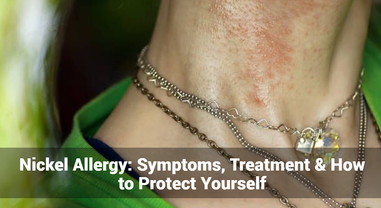
Nickel Allergy: Symptoms, Treatment & How to Protect Yourself

Nickel allergy is a common cause of allergic contact dermatitis, affecting millions of people worldwide. Understanding its symptoms, effective treatments, and preventive measures can significantly improve the quality of life for those affected. This comprehensive guide will delve into everything you need to know about nickel allergy.

What is Nickel Allergy?

Nickel allergy is a hypersensitivity reaction of the immune system to nickel, a metal found in many everyday items. When someone with a nickel allergy comes into contact with nickel-containing objects, their skin may react with an itchy rash, known as allergic contact dermatitis.

What Causes of Nickel Allergy?

Nickel allergy is an immune system response to nickel, a metal commonly found in various everyday items. The exact cause of the allergy involves several factors:

  1. Genetic Predisposition: A family history of allergies, including nickel allergy, can increase the likelihood of developing the condition. Genetics plays a role in how the immune system reacts to allergens.
  2. Prolonged Exposure: Continuous or repeated exposure to nickel can sensitize the immune system, leading to an allergic reaction. This is often seen in individuals who frequently wear nickel-containing jewelry or use nickel-coated objects.
  3. Body Piercings and Jewelry: Nickel is a common component in costume jewelry, earrings, necklaces, and other accessories. Direct and prolonged contact with nickel-containing jewelry, especially through body piercings, can trigger an allergic reaction.
  4. Occupational Exposure: Certain professions, such as construction, hairdressing, and metalworking, involve regular contact with nickel-containing tools and materials. Prolonged occupational exposure can increase the risk of developing a nickel allergy.
  5. Environmental Factors: Nickel is present in various environmental sources, including soil, water, and air. Although less common, exposure to nickel through environmental factors can contribute to sensitization.

Track and Manage your Eczema treatment using a comprehensive Eczema App
Download Eczemaless now


What Are The Common Symptoms of Nickel Allergy?

Nickel allergy symptoms typically manifest within 12 to 48 hours after contact with nickel-containing items. The severity and location of symptoms can vary based on the extent of exposure and individual sensitivity. Common symptoms include:

  1. Itchy Rash: One of the hallmark symptoms of nickel allergy is an itchy rash, known as allergic contact dermatitis. The rash often appears as red, raised bumps on the skin.
  2. Blisters: In more severe cases, the rash may develop into fluid-filled blisters. These blisters can be painful and may ooze or crust over.
  3. Dry, Scaly Skin: Prolonged exposure to nickel can lead to chronic dry, scaly patches of skin. The affected areas may become rough and cracked.
  4. Swelling: The skin around the area of contact with nickel may become swollen and tender.
  5. Burning Sensation: Some individuals experience a burning or stinging sensation on the skin where the nickel made contact.
  6. Eczema: Chronic exposure to nickel can lead to eczema (atopic dermatitis), a more severe form of dermatitis characterized by persistent inflammation, itching, and thickened skin.
  7. Localized Reaction: Symptoms typically occur at the site of contact with nickel-containing items. Common areas affected include earlobes (from earrings), wrists (from watches or bracelets), neck (from necklaces), and waist (from belt buckles).
  8. Systemic Reaction: In rare cases, individuals with severe nickel allergy may experience a systemic reaction, with symptoms spreading beyond the initial contact area. This can include widespread itching, hives, and generalized skin inflammation.

Common Sources of Nickel

Nickel can be found in a variety of everyday items, including:

  • Jewelry (earrings, rings, necklaces)
  • Watches and watchbands
  • Eyeglass frames
  • Coins
  • Zippers, buttons, and belt buckles
  • Cell phones and electronic devices
  • Keys and keychains
  • Kitchen utensils and tools

What Do Doctors Diagnose Of Nickel Allergy?

Diagnosing a nickel allergy involves a combination of medical history assessment, physical examination, and specialized testing. Here’s how dermatologists typically diagnose nickel allergy:

1. Medical History

  • Symptom History: The doctor will ask about your symptoms, including when they started, their severity, and any patterns you’ve noticed.
  • Exposure History: You’ll be asked about your exposure to potential sources of nickel, such as jewelry, clothing fasteners, electronic devices, and occupational exposure.
  • Personal and Family History: Information about your personal and family history of allergies or skin conditions will be gathered.

2. Physical Examination

  • Visual Inspection: The doctor will examine the affected areas of your skin for signs of allergic contact dermatitis, such as redness, itching, blisters, and dry, scaly patches.
  • Pattern Recognition: The doctor will look for characteristic patterns of the rash that are typical of nickel allergy, such as localized reactions at sites of contact with nickel-containing items.

GET IN CONTROL OF YOUR ECZEMA

Use our AI tool to check the severity of Eczema and keep track of your Eczema progress.

Use our AI tool to check the severity of Eczema and keep track of your Eczema progress.

3. Patch Testing

Patch testing is the most reliable method for diagnosing nickel allergy. It involves the following steps:

  • Application of Test Patches: Small amounts of nickel sulfate and other potential allergens are applied to patches, which are then placed on your skin (usually on the back).
  • Observation Period: The patches remain on your skin for 48 hours. You must avoid getting the test area wet during this time.
  • Initial Reading: After 48 hours, the patches are removed, and the doctor examines the skin for any reactions.
  • Final Reading: A follow-up examination is conducted 48-96 hours after the patches are removed to check for delayed reactions.

4. Interpretation of Results

  • Positive Reaction: A positive patch test result for nickel will show localized redness, swelling, and possibly small blisters at the test site, indicating an allergic reaction.
  • Negative Reaction: If there is no reaction at the test site, it suggests that you do not have a nickel allergy.

5. Differential Diagnosis

The doctor may consider other conditions that can cause similar symptoms, such as:

  • Irritant Contact Dermatitis: Caused by direct damage to the skin from irritants.
  • Atopic Dermatitis (Eczema): A chronic skin condition characterized by dry, itchy, and inflamed skin.
  • Other Allergic Reactions: Allergies to other metals or substances.

Treatment Options for Nickel Allergy

While there is no cure for nickel allergy, treatments can help manage symptoms and reduce discomfort. These include:

  1. Topical Corticosteroids: These creams or ointments reduce inflammation and itching.
  2. Oral Antihistamines: Medications like diphenhydramine can help alleviate itching and swelling.
  3. Moisturizers: Keeping the skin hydrated with hypoallergenic moisturizers can prevent dryness and irritation.
  4. Cool Compresses: Applying cool, damp cloths to the affected areas can soothe itching and reduce inflammation.
  5. Avoiding Nickel Exposure: The most effective way to manage nickel allergy is to avoid contact with nickel-containing items.

How to Protect Yourself from Nickel Allergy?

Nickel allergy can be challenging to manage due to the prevalence of nickel in everyday items. However, with some proactive measures, you can significantly reduce your risk of exposure and manage your symptoms effectively. Here’s how to protect yourself from nickel allergy:

  1. Choose Nickel-Free Jewelry: Opt for jewelry made from stainless steel, titanium, platinum, or gold (at least 14 karats).
  2. Use Protective Barriers: Apply clear nail polish or a nickel barrier cream to items that may contain nickel.
  3. Wear Protective Clothing: Cover areas of skin that may come into contact with nickel, such as wearing long sleeves to avoid contact with watchbands or belt buckles.
  4. Switch to Plastic or Wooden Items: Replace metal zippers, buttons, and tools with plastic or wooden alternatives.
  5. Be Mindful of Electronics: Use phone cases and covers to minimize direct contact with nickel-containing electronic devices.
  6. Check Product Labels: Look for items labeled “nickel-free” or “hypoallergenic” when purchasing personal items.

Living with Nickel Allergy

Living with a nickel allergy requires vigilance and proactive management. Here are some additional tips to help you navigate daily life:

  1. Educate Yourself: Learn about the sources of nickel and how to avoid them.
  2. Create a Safe Environment: Identify and remove nickel-containing items from your home and workplace.
  3. Communicate Your Allergy: Inform friends, family, and colleagues about your allergy to ensure they understand your needs and can help you avoid exposure.
  4. Carry Allergy Supplies: Keep antihistamines, corticosteroid creams, and other essential items with you for quick relief in case of accidental exposure.
  5. Regular Dermatologist Visits: Schedule regular check-ups with your dermatologist to monitor your condition and adjust treatment as needed.

Conclusion

Nickel allergy can be a challenging skin condition to manage, but with the right knowledge and precautions, you can significantly reduce your risk of exposure and minimize symptoms. By understanding the causes, recognizing the symptoms, and implementing effective treatments and preventive measures, you can lead a comfortable and healthy life despite your nickel allergy.


Track and Manage your Eczema treatment using a comprehensive Eczema App
Download Eczemaless now

Erythema Infectiosum (Fifth Disease) – Causes, Symptoms, & Treatment

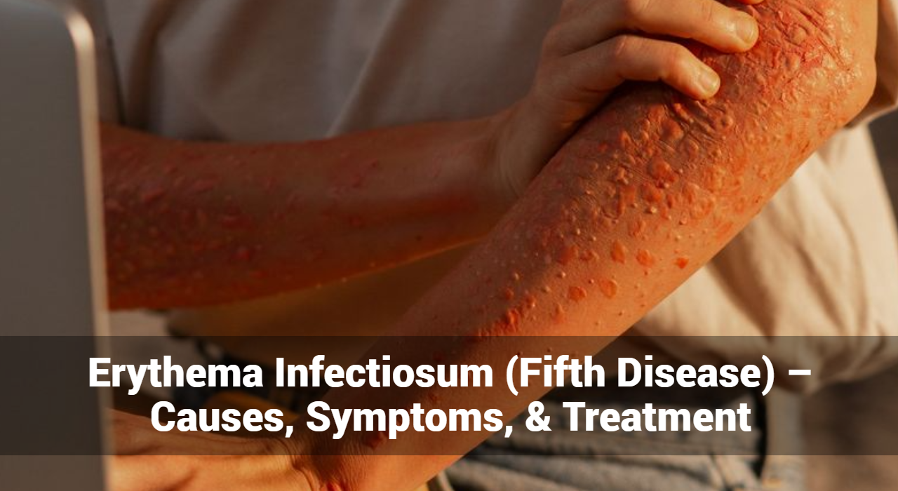
Erythema infectiosum, commonly known as fifth disease, is a highly contagious viral infection primarily affecting children. Recognized by its distinctive “slapped cheek” rash, erythema infectiosum is generally mild but can pose significant risks to certain populations. This article delves into the details of erythema infectiosum, exploring its causes, symptoms, and treatment.

What Is Erythema Infectiosum?

Erythema infectiosum is caused by the human parvovirus B19. It is one of several childhood exanthems (rashes) and is so named because it was historically the fifth disease in a classification of common childhood rashes. The condition is typically mild in healthy children but can have more severe implications for adults and individuals with weakened immune systems.

What Are The Causes of Erythema Infectiosum?

Erythema infectiosum, also known as fifth disease, is a viral infection primarily caused by the human parvovirus B19. This section will delve into the specific causes and contributing factors associated with erythema infectiosum.

Primary Cause: Human Parvovirus B19

Human parvovirus B19 is the sole causative agent of erythema infectiosum. This virus specifically targets and infects red blood cell precursors in the bone marrow, leading to a temporary disruption in red blood cell production. The infection process and subsequent immune response are what primarily lead to the symptoms observed in erythema infectiosum.

Transmission Methods

Erythema infectiosum spreads primarily through the following means:

  1. Respiratory Droplets: The most common transmission route is via respiratory droplets. When an infected person coughs or sneezes, they release tiny droplets containing the virus into the air. These droplets can be inhaled by nearby individuals, leading to infection.
  2. Direct Contact: The virus can also spread through direct contact with respiratory secretions. For example, sharing utensils, drinks, or touching surfaces contaminated with these secretions can facilitate the spread of the virus.
  3. Blood Transmission: Less commonly, human parvovirus B19 can be transmitted through blood or blood products. This can occur through transfusions, organ transplants, or shared needles.
  4. Vertical Transmission: Pregnant women infected with human parvovirus B19 can transmit the virus to their fetus. This vertical transmission can lead to severe complications such as fetal anemia or hydrops fetalis.

Track and Manage your Eczema treatment using a comprehensive Eczema App
Download Eczemaless now


Factors Contributing to the Spread

Several factors contribute to the spread and impact of erythema infectiosum:

  1. Seasonal Variation: Outbreaks of erythema infectiosum are more common in late winter and early spring, although the virus can circulate year-round.
  2. Close Contact Environments: Settings such as schools, daycare centers, and households where individuals are in close proximity to each other facilitate the spread of the virus. Children are particularly susceptible due to their close interactions and developing immune systems.
  3. Immunocompromised Individuals: People with weakened immune systems, including those undergoing chemotherapy, organ transplant recipients, or individuals with HIV, are more susceptible to infection and may experience more severe symptoms.
  4. Chronic Anemia Conditions: Individuals with chronic anemia conditions, such as sickle cell disease or hereditary spherocytosis, are at higher risk for complications if they contract erythema infectiosum. The virus can exacerbate their condition by further disrupting red blood cell production.

Pathogenesis

The pathogenesis of erythema infectiosum involves several stages:

  1. Viral Entry and Replication: The human parvovirus B19 enters the body through the respiratory tract and initially replicates in the nasopharynx.
  2. Viremia: The virus then enters the bloodstream, leading to viremia. During this stage, the infected individual may experience flu-like symptoms.
  3. Immune Response: The body’s immune response to the virus results in the characteristic symptoms of erythema infectiosum, including the distinctive rash and joint pain. The immune response also helps to clear the virus from the body.
  4. Bone Marrow Suppression: Human parvovirus B19 targets red blood cell precursors in the bone marrow, temporarily halting their production. This effect is usually mild and transient in healthy individuals but can be severe in those with pre-existing anemia conditions.

Erythema infectiosum is caused by the human parvovirus B19, which spreads primarily through respiratory droplets, direct contact with infected secretions, blood transmission, and vertical transmission from mother to fetus.

What Are The Symptoms of Erythema Infectiosum?

Erythema infectiosum, commonly known as fifth disease, is a viral infection with a distinctive set of symptoms that evolve through different stages of the illness. Understanding these symptoms helps in early recognition and appropriate management of the condition.

Incubation Period

The incubation period for erythema infectiosum ranges from 4 to 14 days, although it can extend to 21 days in some cases. During this time, the infected person may not exhibit any noticeable symptoms.

Early Symptoms

Initial symptoms are often mild and nonspecific, resembling those of a common cold or flu. These can include:

  • Mild Fever: A low-grade fever is common, typically around 101°F (38.3°C).
  • Headache: Generalized headache.
  • Flu-like Symptoms: Including fatigue, muscle aches, and malaise.
  • Sore Throat: Mild throat irritation or soreness.
  • Runny or Stuffy Nose: Symptoms similar to a mild upper respiratory infection.

Rash Development

The hallmark of erythema infectiosum is its characteristic rash, which progresses through distinct stages:

  1. Slapped Cheek” Rash:
    • Appearance: Bright red rash on both cheeks, giving the appearance of slapped cheeks.
    • Timing: This facial rash typically appears after the early symptoms have subsided and the fever has resolved.
    • Common in Children: This symptom is most prominent in children and less common in adults.
  2. Body Rash:
    • Appearance: A lacy, red rash that can spread from the face to the trunk, arms, and legs.
    • Pattern: The rash has a reticular (net-like) pattern, often described as lacy or “fishnet-like.”
    • Itching: The body rash may be itchy, though the severity of itching varies.
  3. Reappearance:
    • Triggers: The rash can fade and reappear over several weeks, often triggered by factors such as sunlight exposure, heat, exercise, or stress.
    • Fluctuation: The intensity and distribution of the rash can fluctuate, appearing more pronounced under certain conditions.

Joint Pain and Swelling

Joint involvement is more common in adults than in children and can include:

  • Arthralgia: Pain in the joints, commonly affecting the hands, wrists, knees, and ankles.
  • Arthritis: In some cases, there may be swelling and inflammation of the joints, leading to arthritis-like symptoms.
  • Duration: Joint pain and swelling can last from a few days to several weeks or even months, but they typically resolve without long-term damage.

Additional Symptoms

In addition to the classic rash and joint symptoms, erythema infectiosum can present with other signs, especially in adults or those with compromised immune systems:

  • Generalized Fatigue: Persistent tiredness and lack of energy.
  • Gastrointestinal Symptoms: Mild nausea or abdominal discomfort in some cases.
  • Respiratory Symptoms: Cough and mild respiratory distress.


GET IN CONTROL OF YOUR ECZEMA

Use our AI tool to check the severity of Eczema and keep track of your Eczema progress.

Use our AI tool to check the severity of Eczema and keep track of your Eczema progress.


Treatment of Erythema Infectiosum

While it usually resolves on its own without specific antiviral treatment, managing the symptoms can provide relief and prevent complications. Here’s an in-depth look at the treatment strategies for erythema infectiosum.

General Management

The cornerstone of treating erythema infectiosum involves supportive care to alleviate symptoms and ensure comfort. Here are the key approaches:

Symptomatic Relief

  • Fever and Pain: Over-the-counter medications such as acetaminophen (Tylenol) or ibuprofen (Advil, Motrin) can be used to reduce fever and relieve pain, including headache and joint discomfort.
  • Itching: Antihistamines (such as diphenhydramine or cetirizine) may help reduce itching associated with the rash.

Hydration and Rest

  • Fluids: Encourage adequate fluid intake to stay hydrated, especially if the patient has a fever.
  • Rest: Ensure plenty of rest to help the body recover from the viral infection.

Skin Care

  • Moisturizers: Use gentle moisturizers to soothe irritated skin.
  • Avoid Triggers: Minimize exposure to sunlight, heat, and activities that might exacerbate the rash.

Specific Considerations for Different Populations

Children

  • Comfort Measures: Ensuring comfort through appropriate clothing and a cool environment can help manage symptoms.
  • Monitoring: Keep an eye on symptoms and ensure they don’t escalate, particularly if the child has underlying health issues.

Adults

  • Joint Pain Management: Since joint pain and swelling are more common in adults, NSAIDs like ibuprofen can be particularly helpful. In some cases, more potent anti-inflammatory medications may be needed.

Pregnant Women

  • Monitoring: Pregnant women who contract erythema infectiosum should be closely monitored due to the risk of fetal complications. Regular ultrasounds may be recommended to check for signs of fetal distress, such as hydrops fetalis.
  • Medical Consultation: Immediate consultation with a healthcare provider is essential if a pregnant woman is exposed to or develops symptoms of erythema infectiosum.

Immunocompromised Individuals

  • Medical Management: Those with weakened immune systems, such as patients undergoing chemotherapy or those with HIV, may require more intensive medical care. This can include antiviral medications and treatments to support the immune system.
  • Monitoring for Complications: Close monitoring for potential complications, such as severe anemia, is crucial.

Individuals with Chronic Anemia

  • Aplastic Crisis Management: Individuals with health conditions like sickle cell disease or other hemolytic anemias are at risk for aplastic crises. Treatment may include blood transfusions and hospitalization to manage severe anemia.
  • Regular Check-ups: Ongoing monitoring and supportive care to manage their underlying condition and prevent complications.

Preventive Measures

Good Hygiene Practices

  • Hand Washing: Regular and thorough hand washing with soap and water can help prevent the spread of the virus.
  • Respiratory Etiquette: Covering the mouth and nose with a tissue or elbow when coughing or sneezing can reduce the risk of transmission.

Avoiding Exposure

  • Stay Home: Infected individuals, especially children, should stay home from school or daycare during the contagious period to prevent spreading the virus to others.
  • Isolate: Avoid close contact with others, particularly vulnerable groups such as pregnant women and immunocompromised individuals.

When to Seek Medical Attention

While erythema infectiosum is typically mild, certain situations warrant medical attention:

  • Severe Symptoms: Persistent high fever, severe headache, or intense joint pain.
  • Complications: Signs of complications such as difficulty breathing, significant swelling, or prolonged symptoms.
  • Pregnancy: If a pregnant woman is exposed to the virus or develops symptoms.
  • Immunocompromised Individuals: Any signs of severe illness or complications in individuals with weakened immune systems.

Prevention of Erythema Infectiosum

Preventive measures focus on good hygiene and avoiding exposure to the virus:

  • Good Hygiene Practices: Frequent handwashing, avoiding close contact with infected individuals, and proper respiratory etiquette (covering mouth and nose when coughing or sneezing).
  • Avoiding Exposure: Pregnant women and immunocompromised individuals should avoid contact with known cases of erythema infectiosum.

Conclusion

Erythema infectiosum is a common viral infection marked by its characteristic rash and mild flu-like symptoms. While typically mild, it can lead to complications in pregnant women, immunocompromised individuals, and those with chronic anemia. Understanding the causes, symptoms, and treatment options for erythema infectiosum can help manage and prevent the spread of this infection effectively. With proper care and preventive measures, the impact of erythema infectiosum can be minimized, ensuring better health outcomes for affected individuals.


Track and Manage your Eczema treatment using a comprehensive Eczema App
Download Eczemaless now

Treatment & Care for Eczema Infections

Table of Content

Abstract

Eczema also commonly know known as Atopic Dermatitis can become so infected that the person has to go to the hospital and stay over for treatment, this can be very upsetting, especially for youngsters which means missing days of school. Some infections, such as eczema herpeticum (a viral infection), are serious and need medical attention right away, you don’t have any option and in case, if left untreated it may cause sepsis which can be life-threatening.

Some kinds of skin infections can be treated with antibiotics (in the form of tablets, creams, injections or IV drips). Other kinds of skin infections are fungal (such as ringworm) and are treated with antifungal creams or tablets.

It is very obvious that one should immediately visit the physicians for advice to fight back the infection and to find a perfect eczema cure but as it always said that “prevention is better than cure”. Let’s check out what all things can be done to avoid infections and smooth eczema management.

Prevention measure to avoid Eczema Infection

  • It is important to keep your skin as healthy as possible to avoid infection, especially during an Eczema flare. When flares occur, a person should follow the recommended treatment plan to help manage and reduce the flare.
  • If you are suffering from Eczema, avoid contact with anyone who has cold sores. Cold sores are highly infectious. Since the presence of Eczema reduces the immunity against viral infections, eczematous lesions can get infected easily.
  • Frequent hand washing – As we touch surfaces all the time, it is best to wash our hands frequently, especially if they get contaminated with germs.
  • Avoid touching your eczema lesions unnecessarily as you can introduce germs to the rash
  • Avoid scratching – Scratching can damage the skin and break the natural surface barrier for infections. Cut and maintain your nails so that it doesn’t hurt much in case you scratched unknowingly
  • Keep the rashes and skin moisturized well for extra protection.
  • Avoid trigger factors which worsen eczema (Synthetic fabrics, dyes, soaps etc)
  • Follow a healthy diet and avoid foods that you may be sensitive for e.g. nuts and dairy products
  • Keep your skin as clean as possible
  • Children who have eczema should be monitored closely and reminded not to scratch.
  • If flares of Eczema occur, seek treatment early and stick to the recommended treatment plan. The more severe your eczema, it is more prone to infection.
  • Keep your environment clean, free from dust and animal dander
  • Manage your stress – as stress is known to trigger eczema, managing your stress well can reduce flares and thus infections. Practice relaxation techniques, yoga, and meditation.

You can manage your care plan and daily routine by an eczema tool to check what care plan is working for you and stick to it for effective results.

What you can do as a home remedy?

Bath/shower

  • Bath or shower every day to clean the skin.
  • Use warm water and a soft cloth to gently soak and lift off any crusts.
  • Use a soap-free wash e.g. non-ionic cream, aqueous cream, emulsifying ointment. Don’t use soap and bubble baths as these make the skin dry.
  • Antiseptic baths two times a week can help. See bleach bath instructions.

Steroid creams and ointments

  • Apply steroid to all red and itchy skin (active eczema) at-least once a day. Immediately after the bath is best.
  • Use enough to make the skin shiny. Steroid for the face/neck: Steroid for the body/arms/legs:
  • When the skin is no longer red and itchy stop using the steroid but keeps it moisturized. If eczema comes back, start using the steroid again

Moisturizer (emollient)

  • Smooth on lots of moisturizers many times a day to keep the skin soft.
  • Apply all over not just where there is eczema.

Treatment for Eczema Infection

Once the infection breaches your prevention, immediately look for treatment.

On approaching the medical care, the physician may take skin from the site which will be sent for pathology testing. The Microbiological testing of the smear helps to identify the type of infection. The mode of treatment will majorly depend on the result of the test whereas empirical treatment can be started without delay. According to the results of the culture and antibiotic sensitivity testing, treatment can be modified.

  • If the infection is mild an antibiotic cream or ointment will be prescribed. e.g: Neosporin, Polysporin, Fucidine.
  • Sometimes the antibiotic is combined with a steroid. e.g: Betnovate N, Fucicort, Corticosporin.
  • When the infection is widespread, an oral antibiotic will be added. e.g: a course of Flucloxacillin or Co-Amoxyclav to fight the infection better.
  • To infants and children with infected Eczema, oral antibiotics will be given in syrup form, whereas for adults, tablets and capsules are preferred.
  • If the patient is ill with fever and chills, your doctor will admit you and treat the infected Eczema with IV antibiotics.
  • Sometimes steroids can worsen infections. Topical immune-modulators like Protopic ointment and Elidel cream are preferred to steroids by some doctors when treating infected Eczema.
  • Viral infections are treated with oral antiviral medications e.g: Oral Acyclovir for 1 week.
    Sometimes an antiviral cream (Herperax) can be applied topically over the rash. Eczema which is infected with a virus can heal spontaneously with time even without treatment with anti-viral medications. However, if it doesn’t heal seek treatment.
  • If Eczema Herpeticum is severe, hospital admission is required and drugs will be given via a drip.
  • If there is a pain, pain relief can be done by Tylenol (Acetaminophen) or Advil (Ibuprofen). These are also available as over the counter products. Make sure that you adhere to the proper dose and dosage instructions.
  • Treatment of Fungal infections of Eczema – Use of a cream or ointment containing antifungal and steroid combinations.
    e.g: Candacort (Clotrimazole and Hydrocortisone)
    Ecocort (Econazole and Triamcinolone)
    Candid B (Betamethasone and Clotrimazole)
    Once the inflammation is controlled you may be treated with a pure antifungal cream or an ointment. Sometimes your doctor may first control the fungal infection with a pure antifungal cream or ointment rather than a combination.
    e.g: Clotrimazole (Lotrimin), Lamisil (Terbinafin), Tolnaftate
    Once the fungal infection is controlled treatment will follow with usual topical products that control the Eczema rash.
  • Sometimes fungal infections can be widespread especially in immune-compromised patients such as those who suffer from immunity disorders, AIDS, Cancer, etc. Then a course of oral or intravenous antifungal medication will be added depending on severity.


In addition to the specific treatment of infection, the usual treatment for Eczema should also be followed, such as;

Moisturizing the skin well – Moisturize your skin adequately with a good emollient twice a day, especially after a bath, while the skin is still damp. Emollients with minimal fragrance, which are alcohol and paraben-free, are the best. Ingredients in a good moisturizer are glycerol, Dimethicone, aqueous cream, Lanolin oil, Shea butter, Argon oil, cocoa butter, etc. Moisturizers are best when used in the Ointment form rather than a cream form. Choose the best emollient which is suitable for your skin or get a prescription from your doctor which will be the best for you.

Management of itching with an antihistamine – These are also available as over the counter products.
e.g: cetirizine (Alerid, Cetzine), Loritidine (Claritin, Claratyne), fexofenadine (Allegra) or Chlorpheniramine (Piriton) to reduce itching.
As antihistamine products prevent you from the urge of scratching it will help the control of further skin damage and infection.

Wet dressing or bandages to cover and treat eczema rash – This helps to maintain moisture and to prevent further damage to the skin by scratching. However, bandages are preferred once the infection is controlled. Avoid applying bandages when eczema is infected.

When to look back to your physician

  • If you find that the infection is not at all improving after 2–3 days of treatment
  • If your child is missing school due to severe skin infections or not sleeping well because of eczema
  • Once you complete the course and find the symptoms are recurring

GET IN CONTROL OF YOUR ECZEMA

Use our AI tool to check the severity of Eczema and keep track of your Eczema progress.

Use our AI tool to check the severity of Eczema and keep track of your Eczema progress.