What Are Cherry Angiomas? It’s Causes, Symptoms & How To Treat

Cherry angiomas, also known as senile angiomas or Campbell de Morgan spots, are benign skin growths that can appear as small, red or purple spots on the skin. Although they are harmless, their appearance can be concerning to many individuals. In this article, we will explore the causes, symptoms, treatment options, and removal methods associated with cherry angiomas to provide a comprehensive understanding of this common skin condition.

What is a Cherry Angioma?

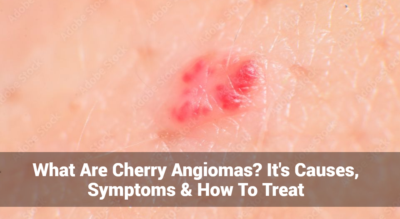
A cherry angioma is a type of vascular lesion that results from the proliferation of small blood vessels (capillaries) in the skin. These growths are usually bright red, but they can also appear blue or purple. They are most commonly found on the trunk, arms, and shoulders, but they can develop anywhere on the body.

Characteristics of Cherry Angiomas

  • Color: Typically bright red but may appear purple or blue.
  • Size: Usually small, ranging from a pinpoint to about a quarter of an inch in diameter.
  • Texture: Smooth and dome-shaped, but they can also be flat.
  • Location: Commonly found on the torso, arms, and neck, but they can occur anywhere on the body.

Track and Manage your Eczema treatment using a comprehensive Eczema App
Download Eczemaless now


What Causes of Cherry Angiomas?

Cherry angiomas are benign growths that typically appear as small, red, or purple spots on the skin. While the exact cause of cherry angiomas is not fully understood, several factors have been identified that may contribute to their development. Here’s a closer look at these potential causes:

  • Family History: Cherry angiomas tend to run in families, suggesting a genetic predisposition. If your parents or siblings have cherry angiomas, you may be more likely to develop them as well.
  • Incidence Increases with Age: The likelihood of developing cherry angiomas increases with age, particularly in individuals over 30. This age-related prevalence suggests that as the skin ages, its structure and blood vessels may become more susceptible to the formation of these growths.
  • Pregnancy and Hormonal Fluctuations: Changes in hormone levels, such as during pregnancy, can trigger the development of cherry angiomas. Hormonal shifts may affect blood vessel growth and skin changes, leading to the appearance of these angiomas.
  • Chemical Exposure: While specific environmental triggers have not been conclusively identified, some studies suggest that exposure to certain chemicals or irritants could contribute to the development of cherry angiomas. For instance, prolonged exposure to solvents or other chemical agents may play a role.
  • Injury or Irritation: Some individuals report that cherry angiomas developed after skin trauma or irritation. While the connection isn’t definitively established, physical damage to the skin could potentially trigger the growth of these vascular lesions.
  • Certain Health Conditions: In some cases, cherry angiomas may be associated with underlying medical conditions, such as hormonal disorders. However, this is less common, and the majority of cherry angiomas occur in otherwise healthy individuals.

GET IN CONTROL OF YOUR ECZEMA

Use our AI tool to check the severity of Eczema and keep track of your Eczema progress.

Use our AI tool to check the severity of Eczema and keep track of your Eczema progress.

What Are The Symptoms of Cherry Angiomas?

While they typically do not cause significant symptoms, there are several characteristics and signs associated with cherry angiomas that individuals may observe. Here are the main symptoms:

1. Appearance

  • Color: Cherry angiomas are usually bright red but can also appear purple or blue.
  • Size: They typically range from a few millimeters to about a quarter of an inch in diameter.
  • Shape: Most cherry angiomas have a smooth, dome-shaped surface, although some may be flat or slightly raised.
  • Texture: The surface is usually smooth, but they can also be rough or scabby in some cases.

2. Location

  • Common Areas: Cherry angiomas commonly appear on the trunk, arms, shoulders, and neck but can develop anywhere on the body, including the face and scalp.

3. Bleeding

  • Injury: Cherry angiomas can bleed if they are accidentally scraped or cut. While they usually do not cause pain, bleeding can be a concern if the growth is injured.
  • Spontaneous Bleeding: In rare cases, individuals may experience spontaneous bleeding from a cherry angioma, which may require medical attention.

4. Change in Size or Color

  • Growth Over Time: Cherry angiomas may gradually increase in size or number over time. While this is common, any sudden changes in size, shape, or color should be evaluated by a healthcare professional.
  • Color Changes: Some individuals may notice a darkening of the angioma over time.

5. Itching or Irritation (Rare)

  • Mild Discomfort: Although cherry angiomas are typically painless, some individuals may experience mild itching or irritation, especially if the angioma is located in an area that experiences friction or irritation from clothing.

6. Psychological Impact

Cosmetic Concerns: The appearance of cherry angiomas can lead to self-consciousness or concerns about one’s appearance, especially if they are numerous or located on visible areas of the body.

Diagnosis of Cherry Angiomas

Diagnosing a cherry angioma typically involves a physical examination by a healthcare provider or consult a dermatologist. In most cases, no additional testing is necessary. However, if there is any doubt about the diagnosis, a biopsy may be performed to ensure that the growth is benign.

GET IN CONTROL OF YOUR ECZEMA

Use our AI tool to check the severity of Eczema and keep track of your Eczema progress.

Use our AI tool to check the severity of Eczema and keep track of your Eczema progress.

What Are The Treatment Options for Cherry Angiomas?

While cherry angiomas are harmless and do not require treatment, some individuals choose to have them removed for cosmetic reasons. Several treatment options are available:

1. Laser Therapy: Laser therapy is a common method for removing cherry angiomas. A concentrated beam of light targets the blood vessels, causing them to collapse and fade. This treatment is quick and often requires little to no downtime.

2. Cryotherapy: Cryotherapy involves freezing the cherry angioma with liquid nitrogen. This process destroys the abnormal blood vessels, causing the growth to fall off after a few days.

3. Electrosurgery: In this method, a high-frequency electrical current is used to burn off the cherry angioma. This treatment can be effective for larger growths.

4. Surgical Excision: For larger or multiple cherry angiomas, surgical excision may be performed. This involves cutting out the growth with a scalpel, followed by suturing the skin. This method is often reserved for cases where other treatments are not suitable.

Home Remedies for Cherry Angiomas

While medical treatments are the most effective way to remove cherry angiomas, some home remedies may help improve their appearance or prevent new ones from forming:

  • Apple Cider Vinegar: Applying apple cider vinegar to the angioma may help reduce its size over time.
  • Tea Tree Oil: This essential oil is believed to have properties that may help reduce the appearance of cherry angiomas when applied topically.
  • Healthy Lifestyle: Maintaining a balanced diet, staying hydrated, and protecting the skin from excessive sun exposure can promote overall skin health.

When to See a Doctor

While cherry angiomas are usually harmless, individuals should seek medical advice if they notice any of the following:

  • Rapid Changes: Sudden changes in size, shape, or color of the angioma.
  • Excessive Bleeding: Bleeding that does not stop or occurs frequently.
  • New Growths: The development of new skin growths that differ in appearance from existing cherry angiomas.
  • Associated Symptoms: Any discomfort, pain, or unusual symptoms associated with the angioma.

Prevention of Cherry Angiomas

Since the exact cause of cherry angiomas is not fully understood, there is no guaranteed way to prevent them. However, some general tips for maintaining healthy skin include:

  • Sun Protection: Use sunscreen to protect your skin from harmful UV rays.
  • Healthy Diet: A diet rich in fruits, vegetables, and antioxidants may contribute to skin health.
  • Avoid Harsh Chemicals: Limit exposure to chemicals and irritants that may affect skin health.

Conclusion

Cherry angiomas are common, benign skin growths that can occur as people age. While they are generally harmless, many individuals seek treatment for cosmetic reasons. Understanding the causes, symptoms, and available treatment options can help individuals make informed decisions about their skin health. If you have concerns about cherry angiomas or any changes in your skin, it is always best to consult a healthcare professional for proper evaluation and advice.

By staying informed about cherry angiomas and maintaining good skin care practices, individuals can navigate this common skin condition with confidence.


Track and Manage your Eczema treatment using a comprehensive Eczema App
Download Eczemaless now

Adult Acne: Types, Causes, Symptoms, Treatment & Prevention

Adult acne is a frustrating skin condition that affects many individuals beyond their teenage years. While often associated with puberty, it can persist well into adulthood, impacting self-esteem and overall skin health. In this comprehensive guide, we will explore the types, causes, symptoms, treatment options, and prevention strategies for adult acne.

Understanding Adult Acne

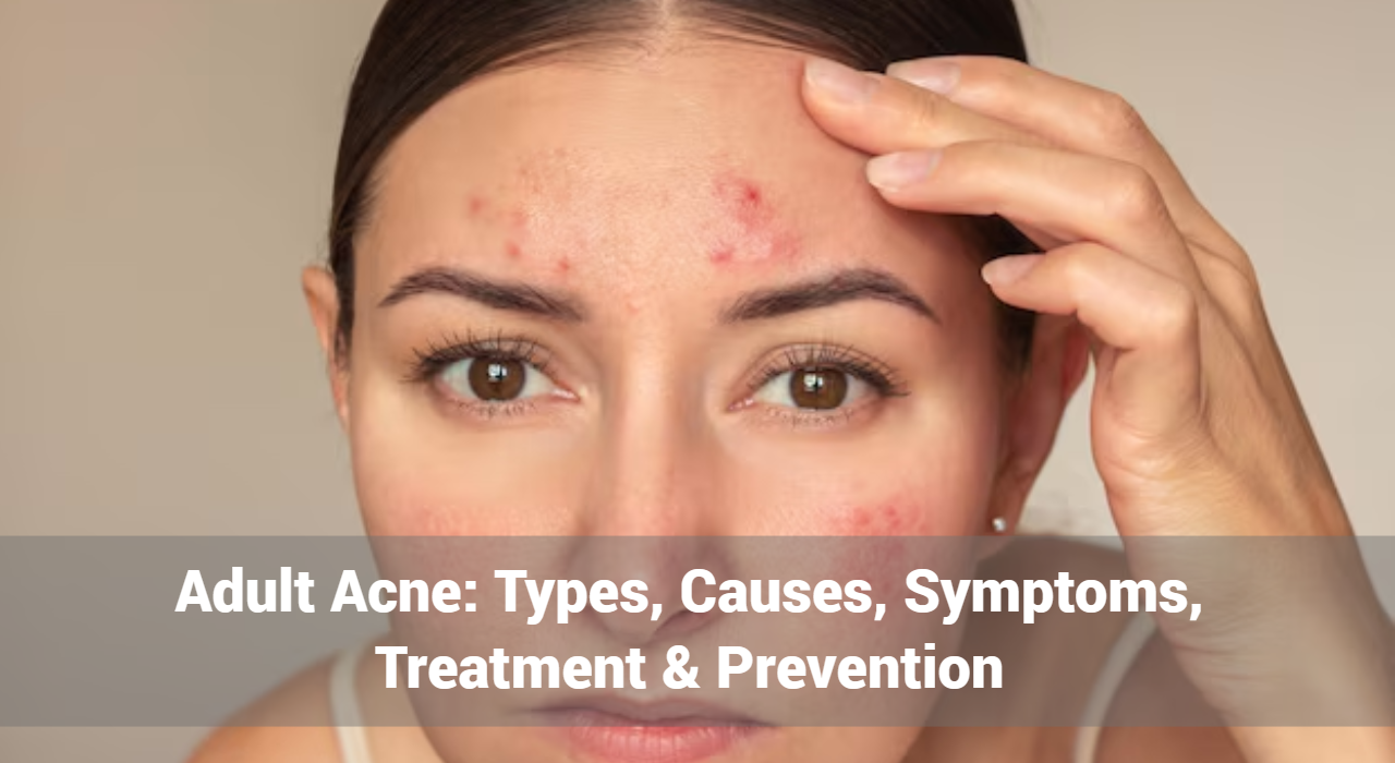
Adult acne refers to acne that appears after the age of 25. It can manifest in various forms, such as pimples, blackheads, and cysts, and can occur on different areas of the body, though it primarily affects the face, neck, and back. Recognizing the specific characteristics of adult acne is essential for effective treatment.

Common Different Types of Adult Acne

  1. Comedonal Acne: This type includes blackheads and whiteheads. Comedones occur when hair follicles become clogged with oil and dead skin cells.
  2. Inflammatory Acne: Characterized by red, swollen bumps, this type includes papules and pustules. Inflammatory acne is often more painful and can lead to scarring.
  3. Nodular Acne: Larger, painful lumps that form deep within the skin. Nodular acne is more severe and requires targeted treatment.
  4. Cystic Acne: This severe form of acne consists of painful, pus-filled cysts. Cystic acne can lead to significant scarring and often necessitates medical intervention.

Track and Manage your Eczema treatment using a comprehensive Eczema App
Download Eczemaless now


What Causes of Adult Acne?

Understanding the underlying causes of adult acne is crucial for effective management. Some of the most common triggers include:

  • Hormonal Fluctuations: Changes in hormones, particularly during menstruation, pregnancy, or menopause, can lead to increased oil production and clogged pores.
  • Stress: High-stress levels can trigger hormonal changes that exacerbate acne. Stress leads to increased production of cortisol, which in turn stimulates oil glands.
  • Diet: Certain foods, especially those high in sugar and dairy, have been linked to acne flare-ups. A diet rich in processed foods can also contribute to inflammation.
  • Medications: Some medications, such as corticosteroids, certain birth control pills, and medications containing lithium, can cause acne as a side effect.
  • Skin Care Products: Using heavy, comedogenic products can clog pores. It’s essential to choose non-comedogenic formulations to minimize the risk of breakouts.
  • Environmental Factors: Pollution and humidity can contribute to acne by irritating the skin and clogging pores.

What Are The Symptoms of Adult Acne?

Adult acne can present in various forms, and recognizing its symptoms is crucial for effective treatment. Here are the common symptoms associated with adult acne:

  1. Pimples: Red, inflamed bumps that can be painful. They often have a white or yellow pus-filled center.
  2. Blackheads: Open comedones that appear dark due to oxidized sebum. They are commonly found on the nose and forehead.
  3. Whiteheads: Closed comedones that remain beneath the skin’s surface, appearing as small, flesh-colored bumps.
  4. Cysts: Large, painful lumps filled with pus that form deep within the skin. They can be particularly severe and may lead to scarring.
  5. Papules: Small, red, raised bumps that are usually tender to the touch. They are often a sign of inflammation.
  6. Pustules: Similar to pimples but are filled with pus, often appearing red around the base.
  7. Nodules: Hard, painful lumps that form under the skin. Nodular acne can be severe and requires medical treatment.
  8. Scarring: Over time, untreated acne can lead to various types of scarring, including pitted or raised scars, which can affect skin texture.
  9. Hyperpigmentation: Dark spots or discoloration may develop after acne lesions heal, particularly in individuals with darker skin tones.

Recognizing these symptoms early can help in seeking appropriate treatment and minimizing the risk of scarring and further breakouts. If you experience persistent or severe symptoms, it’s advisable to consult a dermatologist.


GET IN CONTROL OF YOUR ECZEMA

Use our AI tool to check the severity of Eczema and keep track of your Eczema progress.

Use our AI tool to check the severity of Eczema and keep track of your Eczema progress.


Treatment Options for Adult Acne

Effective treatment for adult acne often requires a combination of approaches tailored to individual needs. Here are some commonly recommended options:

1. Topical Treatments

  • Benzoyl Peroxide: Helps kill bacteria and reduce inflammation.
  • Salicylic Acid: A beta-hydroxy acid that helps exfoliate the skin and unclog pores.
  • Retinoids: Vitamin A derivatives that promote cell turnover and prevent clogged pores. Prescription options are often more effective.
  • Antibiotics: Topical antibiotics can reduce bacteria and inflammation in moderate cases.

2. Oral Medications

  • Hormonal Therapy: Birth control pills can regulate hormonal fluctuations that lead to acne.
  • Antibiotics: Oral antibiotics may be prescribed for moderate to severe inflammatory acne.
  • Isotretinoin: A potent medication for severe acne, typically reserved for cases that do not respond to other treatments.

3. Professional Treatments

  • Chemical Peels: Help exfoliate the skin and reduce the appearance of scars and active breakouts.
  • Laser Therapy: Targets acne lesions and helps reduce inflammation and redness.
  • Microneedling: Promotes collagen production to improve skin texture and reduce scars.

Prevention Strategies for Adult Acne

Preventing adult acne requires a multifaceted approach that addresses both skincare and lifestyle factors:

  • Consistent Skincare Routine: Use a gentle cleanser twice a day and follow with non-comedogenic moisturizers and treatments.
  • Balanced Diet: Focus on a diet rich in fruits, vegetables, and whole grains while minimizing sugar and processed foods.
  • Hydration: Drink plenty of water to keep your skin hydrated and support overall health.
  • Stress Management: Engage in activities such as yoga, meditation, or regular exercise to help manage stress levels.
  • Avoid Picking: Resist the urge to pick at or pop pimples, as this can lead to scarring and further inflammation.
  • Regular Dermatologist Visits: Regular check-ups consult a dermatologist can help monitor your skin and adjust treatments as necessary.

Conclusion

Adult acne is a common yet often misunderstood condition. By understanding the types, causes, symptoms, and effective treatment options, individuals can take proactive steps to manage their acne effectively. With the right combination of skincare, lifestyle adjustments, and professional guidance, achieving clear, healthy skin is possible. If you’re struggling with adult acne, don’t hesitate to consult a dermatologist to find the best personalized treatment plan for you. Remember, clear skin is within reach!


Track and Manage your Eczema treatment using a comprehensive Eczema App
Download Eczemaless now


How to Relieve Winter Itch: Causes, Symptoms, and Treatment

Winter can be a beautiful season, but it also brings with it an uncomfortable condition known as winter itch. As the temperatures drop and the air becomes dry, many people experience itchy, flaky skin that can lead to irritation and discomfort. In this comprehensive guide, we’ll explore the causes, symptoms, and effective treatments for winter itch, along with preventive measures to keep your skin healthy during the colder months.

What is Winter Itch?

Winter itch, medically referred to as xerotic eczema, is a skin condition characterized by dry, itchy skin that typically worsens in winter. The lack of humidity and the effects of indoor heating can strip the skin of its natural moisture, leading to various uncomfortable symptoms.

What Causes of Winter Itch?

Understanding the underlying causes of winter itch is essential for effective treatment and prevention. Here are the primary factors contributing to this condition:

1. Low Humidity Levels: During winter, outdoor air holds less moisture, which translates to drier skin. This lack of humidity is a major factor in the development of winter itch.

2. Indoor Heating: While central heating keeps us warm, it also reduces indoor humidity, exacerbating skin dryness. The combination of cold air outside and dry air indoors creates a perfect storm for winter itch.

3. Hot Showers: Although a hot shower may feel good in the winter, prolonged exposure to hot water can strip the skin of its natural oils, leading to increased dryness and irritation.

4. Harsh Soaps and Detergents: Many soaps, shampoos, and laundry detergents contain ingredients that can irritate the skin. These products often have harsh chemicals that strip the skin of moisture.

5. Irritating Fabrics: Certain materials, such as wool or synthetic fibers, can irritate sensitive skin, leading to increased itching and discomfort during winter months.

6. Skin Conditions: Individuals with pre-existing skin conditions, such as eczema or psoriasis, may find their symptoms worsen in winter due to the dryness.


Track and Manage your Eczema treatment using a comprehensive Eczema App
Download Eczemaless now


What Are The Common Symptoms of Winter Itch?

Winter itch, or xerotic eczema, presents a range of symptoms that can vary in severity. Here are the common symptoms associated with this condition:

  1. Dry, Flaky Skin: The skin often appears rough and may have visible flakes, resembling dry patches.
  2. Intense Itching: One of the hallmark symptoms is a persistent urge to scratch, which can be particularly bothersome at night.
  3. Redness and Inflammation: Affected areas may become red and inflamed due to irritation and scratching.
  4. Cracking and Scaling: Severe dryness can lead to cracks in the skin, which may bleed or become painful.
  5. Rough Texture: The skin may feel coarse or uneven to the touch.
  6. Sensitive Skin: Affected areas might become more sensitive, reacting to environmental factors like temperature changes or certain fabrics.
  7. Skin Tightness: Individuals may experience a feeling of tightness or discomfort, especially after bathing or exposure to cold air.

What Are The Treatment Options for Winter Itch?

Finding relief from winter itch involves a combination of moisturizing strategies, lifestyle changes, and potential medical treatments. Here’s a detailed approach:

1. Regular Moisturization

Moisturizers are your best defense against winter itch. Look for thick creams or ointments that contain ingredients such as:

  • Ceramides: Help restore the skin barrier.
  • Hyaluronic Acid: Draws moisture into the skin.
  • Glycerin: A humectant that attracts water.

Apply moisturizer immediately after bathing and throughout the day as needed.

2. Modify Bathing Habits

To minimize skin dryness:

  • Opt for lukewarm showers instead of hot ones.
  • Limit shower time to 10-15 minutes.
  • Use gentle, hydrating cleansers that are free of fragrances and harsh chemicals.


GET IN CONTROL OF YOUR ECZEMA

Use our AI tool to check the severity of Eczema and keep track of your Eczema progress.

Use our AI tool to check the severity of Eczema and keep track of your Eczema progress.


3. Increase Humidity

Using a humidifier can significantly improve indoor air quality and moisture levels. Aim to keep indoor humidity levels between 30-50%.

4. Choose Appropriate Clothing

Select soft, breathable fabrics like cotton that are less likely to irritate your skin. Avoid wool and synthetic fibers that can exacerbate itching.

5. Avoid Scratching

While it may be tempting to scratch, doing so can worsen irritation and lead to infections. Keep nails short and consider wearing cotton gloves at night to prevent scratching during sleep.

6. Use Over-the-Counter Remedies

Over-the-counter products can provide relief:

  • Antihistamines: Help manage itching, especially at night.
  • Hydrocortisone Cream: A mild topical steroid that reduces inflammation and itching.

7. Consult a Dermatologist

If winter itch persists despite home treatments, consult a dermatologist. They may prescribe stronger medications or topical treatments tailored to your specific needs.

Prevention Tips for Winter Itch

Preventing winter itch is often easier than treating it. Here are some effective strategies:

1. Stay Hydrated: Drink plenty of water to help maintain skin hydration from within.

2. Follow a Balanced Diet: Incorporate foods rich in omega-3 fatty acids, such as fatty fish, flaxseeds, and walnuts, to support skin health.

3. Use Gentle Skin Care Products: Opt for fragrance-free and hypoallergenic skincare products to minimize irritation.

4. Maintain a Consistent Moisturizing Routine: Make moisturizing a daily habit, especially after bathing or washing hands.

5. Limit Exposure to Cold Air: When outdoors, cover your skin with warm clothing to protect it from the harsh winter elements.

Conclusion

Winter itch can be a frustrating and uncomfortable condition, but understanding its causes, symptoms, and treatment options can help you find relief. By incorporating effective skincare practices and preventive measures, you can keep your skin healthy and itch-free throughout the winter months. If symptoms persist, don’t hesitate to seek professional advice to ensure your skin receives the care it needs. Embrace the beauty of winter without the discomfort of winter itch!


Track and Manage your Eczema treatment using a comprehensive Eczema App
Download Eczemaless now


What Is Kaposi Sarcoma? It’s Symptoms, Causes & Treatment

Kaposi Sarcoma (KS) is a type of cancer that originates in the cells lining the blood vessels and lymphatic vessels. This disease is known for causing abnormal growth of these vessels, resulting in various symptoms and health complications. Although Kaposi Sarcoma is relatively rare, understanding its symptoms, causes, and treatment options is crucial for effective management and better patient outcomes. This comprehensive guide aims to shed light on Kaposi Sarcoma, providing detailed insights into its various aspects.

What is Kaposi Sarcoma?

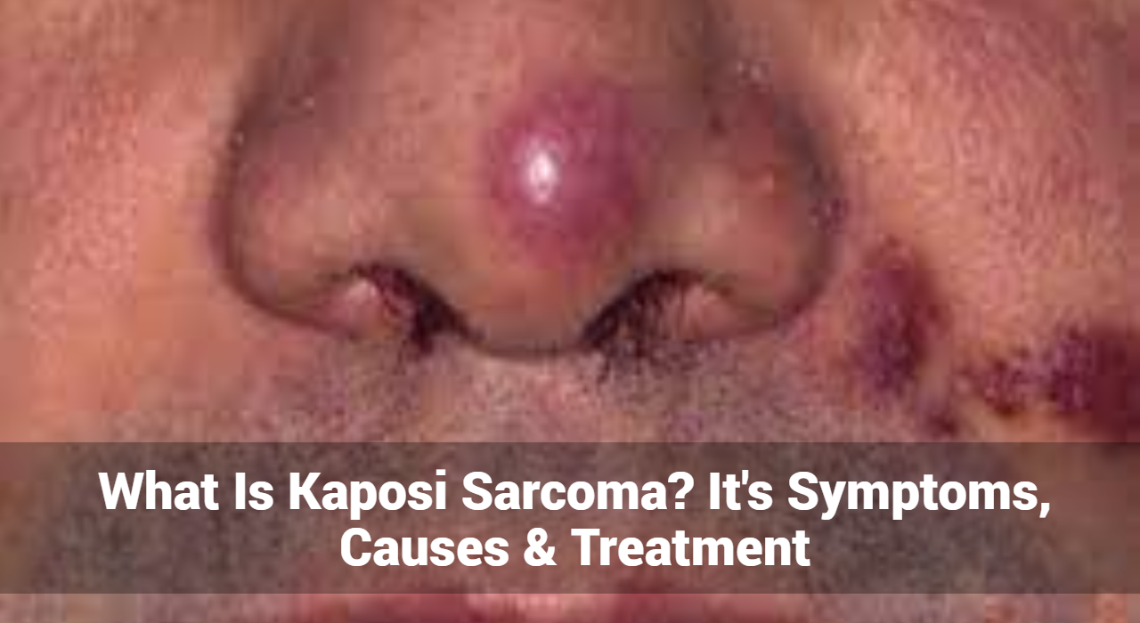
Kaposi Sarcoma is a malignancy that primarily affects the skin but can also involve mucous membranes, lymph nodes, and internal organs such as the lungs, liver, and gastrointestinal tract. The disease is characterized by the formation of reddish-purple or brownish lesions or spots, which may appear on the skin or mucous membranes. KS is often linked with immune system deficiencies and has different forms, including classic, endemic, epidemic (AIDS-related), and iatrogenic.

Different Types of Kaposi Sarcoma

  1. Classic Kaposi Sarcoma: Typically affects older men of Mediterranean or Eastern European descent. It generally progresses slowly and is often limited to the skin.
  2. Endemic Kaposi Sarcoma: Common in certain regions of Africa, particularly among younger individuals. It can be more aggressive compared to the classic form and may involve internal organs.
  3. Epidemic (AIDS-related) Kaposi Sarcoma: Associated with human immunodeficiency virus (HIV) infection and Acquired Immunodeficiency Syndrome (AIDS). This form tends to be more aggressive and can rapidly progress.
  4. Iatrogenic Kaposi Sarcoma: Linked to immunosuppressive therapy, often prescribed for organ transplant recipients. It usually appears when the immune system is significantly weakened.

Track and Manage your Eczema treatment using a comprehensive Eczema App
Download Eczemaless now


What Are The Symptoms of Kaposi Sarcoma?

The symptoms of Kaposi Sarcoma can vary based on the type and location of the lesions. Common symptoms include:

  • Skin Lesions: Red, purple, or brown spots or patches that may appear on the skin, particularly on the legs, feet, or face. These lesions can be flat or raised and may be asymptomatic or cause discomfort.
  • Mucous Membrane Lesions: Lesions may appear in the mouth, throat, or other mucosal surfaces, leading to difficulty swallowing or speaking.
  • Swollen Lymph Nodes: Lymphadenopathy can occur if KS involves the lymphatic system.
  • Gastrointestinal Symptoms: Abdominal pain, nausea, vomiting, and bleeding if internal organs are affected.
  • Respiratory Symptoms: Cough, shortness of breath, and chest pain if the lungs are involved.
  • Systemic Symptoms: Weight loss, fever, and night sweats may occur in more advanced cases.

What Causes of Kaposi Sarcoma?

Kaposi Sarcoma is caused by infection with Human Herpesvirus 8 (HHV-8), also known as Kaposi’s Sarcoma-associated Herpesvirus (KSHV). The virus is primarily spread through sexual contact and is prevalent among individuals with weakened immune systems. The risk factors for developing KS include:

  • Immunosuppression: Individuals with compromised immune systems, such as those with HIV/AIDS or those undergoing immunosuppressive therapy, are at higher risk.
  • HIV/AIDS: KS is strongly associated with HIV infection. The presence of HHV-8 in conjunction with HIV greatly increases the likelihood of developing KS.
  • Age and Gender: Classic KS is more common in older men, while epidemic KS predominantly affects younger males.
  • Geographic Location: Endemic KS is more prevalent in sub-Saharan Africa.

Diagnosis of Kaposi Sarcoma

Diagnosis of Kaposi Sarcoma typically involves a combination of clinical examination and diagnostic tests, including:

  • Physical Examination: A detailed assessment of the lesions and symptoms.
  • Biopsy: A sample of the affected tissue is examined microscopically to confirm the presence of KS.
  • Imaging Studies: CT scans, MRI, or X-rays may be used to assess the extent of internal organ involvement.
  • Blood Tests: To evaluate overall health and detect any signs of systemic involvement.


GET IN CONTROL OF YOUR ECZEMA

Use our AI tool to check the severity of Eczema and keep track of your Eczema progress.

Use our AI tool to check the severity of Eczema and keep track of your Eczema progress.


What Are The Treatment Options of Kaposi Sarcoma?

The treatment of Kaposi Sarcoma depends on the type, stage, and location of the disease, as well as the overall health of the patient. Options include:

  • Antiretroviral Therapy (ART): For AIDS-related KS, managing HIV with ART can lead to significant improvement in KS lesions and overall health.
  • Chemotherapy: Systemic chemotherapy is used for more advanced or widespread KS. Agents like liposomal doxorubicin and paclitaxel are commonly used.
  • Radiation Therapy: Effective for localized KS lesions, particularly those causing pain or discomfort.
  • Immunotherapy: Treatments such as interferon-alpha may be used to boost the immune system and target KS cells.
  • Surgical Treatment: For isolated lesions, surgical removal may be an option, especially if they cause significant symptoms or cosmetic concerns.
  • Targeted Therapy: Newer approaches that specifically target cancer cells or their growth mechanisms are being explored in clinical trials.

Prognosis and Outlook

The prognosis of Kaposi Sarcoma varies widely depending on the type, extent of disease, and response to treatment. In general:

  • Classic KS: Tends to have a slower progression and a better prognosis if limited to the skin.
  • Endemic KS: May be more aggressive, with a variable prognosis depending on internal organ involvement.
  • Epidemic KS: Prognosis improves significantly with effective antiretroviral therapy for HIV.
  • Iatrogenic KS: May improve with adjustments in immunosuppressive therapy.

Conclusion

Kaposi Sarcoma is a complex disease with various forms and presentations. Early detection and tailored treatment strategies are essential for improving outcomes and managing symptoms effectively. Individuals at higher risk, such as those with HIV/AIDS or those undergoing immunosuppressive therapy, should be vigilant and consult a dermatologist or healthcare professional for regular monitoring and management.

By understanding Kaposi Sarcoma’s symptoms, causes, and treatment options, patients and caregivers can make informed decisions and work towards better management of this challenging condition. If you suspect you may have symptoms of Kaposi Sarcoma or are at risk, seek medical advice promptly for appropriate evaluation and care.


Track and Manage your Eczema treatment using a comprehensive Eczema App
Download Eczemaless now


What Is Erythrasma: It’s Symptoms, Causes, And How To Treat

Erythrasma is a common but often under-recognized skin condition that affects many people worldwide. This article will provide a comprehensive overview of erythrasma, including its symptoms, causes, and treatment options, to help you better understand and manage this condition.

What is Erythrasma?

Erythrasma is a chronic skin infection caused by the bacteria Corynebacterium minutissimum. It predominantly affects the skin folds and is characterized by reddish-brown patches with well-defined edges. The condition is often mistaken for other skin disorders, such as fungal infections or eczema, making accurate diagnosis crucial for effective treatment.

Key Characteristics of Erythrasma:

  • Color: The lesions often appear as reddish-brown or pinkish-brown patches.
  • Location: Commonly affects areas where skin folds, such as the armpits, groin, and under the breasts.
  • Appearance: The patches are usually flat, well-defined, and may have a slight scaly texture.
  • Symptoms: Itching is generally mild or absent, which helps differentiate it from other similar conditions.

Track and Manage your Eczema treatment using a comprehensive Eczema App
Download Eczemaless now

What Are The Symptoms of Erythrasma?

Erythrasma is characterized by a range of specific symptoms that help in identifying the condition. Here’s a closer look at what to watch for:

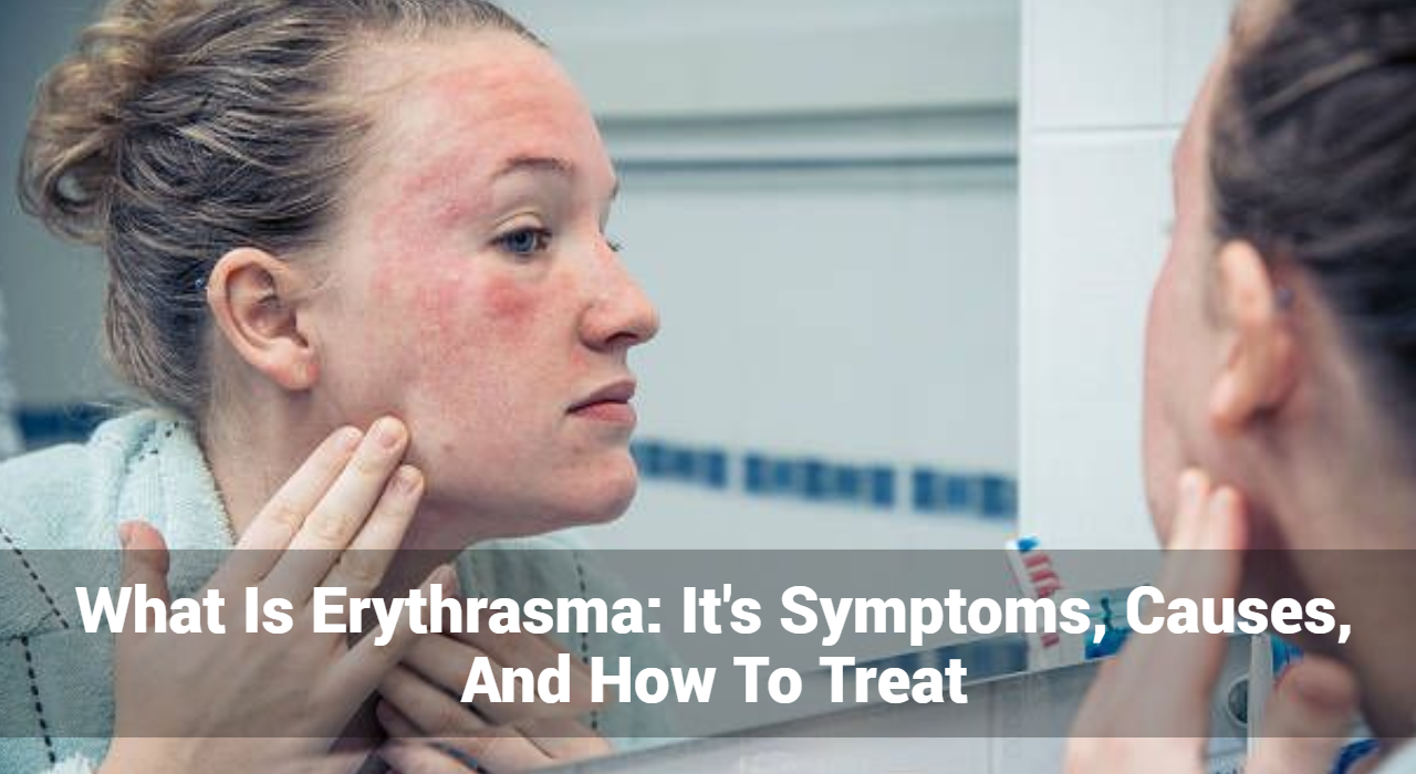
  • Appearance: The hallmark symptom of erythrasma is the development of reddish-brown or pinkish-brown patches on the skin.
  • Edges: These patches are well-defined with clear boundaries, making them easily distinguishable from surrounding skin.
  • Skin Folds: Erythrasma predominantly affects areas where skin folds or where there is friction and moisture. Common sites include:
    • Armpits (axillae)
    • Groin
    • Under the breasts (in women)
    • Between the toes
  • Other Areas: It can occasionally occur in other skin folds or less typical locations.
  • Itching: While itching is not a primary feature, some individuals may experience mild itching or discomfort. This is generally less severe compared to other skin conditions.
  • Discomfort: A slight burning sensation or tenderness may be present, especially if there is additional irritation or secondary infection.
  • Texture: The patches may have a slightly scaly or dry surface, though this is often minimal. The texture can vary, and in some cases, the skin may appear smooth.
  • Inflammation: Erythrasma generally does not cause significant inflammation, swelling, or severe redness. The patches are typically stable and do not lead to systemic symptoms.
  • Possible Odor: In some cases, especially in warm and humid environments, a mild odor may be noticed. This is due to bacterial activity in moist areas.

What Causes of Erythrasma And Risk Factors?

Erythrasma is caused by an overgrowth of the bacterium Corynebacterium minutissimum. This bacterium is normally present on the skin but can proliferate under certain conditions. Here are the primary causes and risk factors associated with erythrasma:

  • Corynebacterium minutissimum: The primary cause of erythrasma is an overgrowth of Corynebacterium minutissimum. This bacterium is part of the normal skin flora but can cause infection when conditions are favorable.
  • Warm and Humid Conditions: Erythrasma is more likely to develop in areas where moisture and warmth are prevalent. Skin folds provide an ideal environment for bacterial growth due to trapped heat and sweat.
  • Inadequate Cleaning: Insufficient washing or cleansing of skin folds can lead to the accumulation of sweat and bacteria, increasing the risk of erythrasma.
  • Diabetes: Individuals with diabetes may be more susceptible to erythrasma due to the impact of high blood sugar levels on skin health.
  • Obesity: Excess weight can create additional skin folds and increase moisture, making the skin more prone to bacterial infections.
  • Weakened Immune System: A compromised immune system may increase the risk of developing erythrasma as it affects the body’s ability to control bacterial growth.
  • Repetitive Friction: Areas subjected to repeated friction, such as from tight clothing, can be more vulnerable to erythrasma. This is especially true in skin folds where friction and moisture can promote bacterial overgrowth.
  • Genetic Predisposition: While not a primary cause, some individuals may be genetically predisposed to skin conditions that could make them more susceptible to erythrasma.
  • Environmental Factors: Exposure to environmental conditions that promote moisture and heat, such as hot and humid climates, can increase the likelihood of developing erythrasma.

Diagnosis of Erythrasma

Proper diagnosis of erythrasma often involves a combination of clinical evaluation and laboratory tests. Dermatologists typically diagnose erythrasma based on the appearance of the skin lesions and their location. To confirm the diagnosis, a few additional tests may be used:

  1. Wood’s Lamp Examination: Erythrasma lesions often fluoresce a coral-red color under a Wood’s lamp, which is a handheld UV light.
  2. Skin Scrapings: In some cases, skin scrapings are examined under a microscope to rule out fungal infections.
  3. Culture Tests: Culturing a sample from the affected area can help identify Corynebacterium minutissimum.


GET IN CONTROL OF YOUR ECZEMA

Use our AI tool to check the severity of Eczema and keep track of your Eczema progress.

Use our AI tool to check the severity of Eczema and keep track of your Eczema progress.


What Are The Treatment Options of Erythrasma?

Treatment of erythrasma typically involves a combination of topical and systemic therapies. The goal is to eliminate the bacteria and alleviate symptoms. Here are the most common treatment options:

1. Topical Antibiotics

Topical antibiotics are often the first line of treatment. They include:

  • Erythromycin: Applied directly to the affected areas to combat the bacterial infection.
  • Clindamycin: Another effective topical antibiotic that targets Corynebacterium minutissimum.

2. Oral Antibiotics

For more extensive or resistant cases, oral antibiotics may be necessary:

  • Doxycycline: A broad-spectrum antibiotic that can effectively treat erythrasma.
  • Tetracycline: Another option that may be used for more severe cases.

3. Good Hygiene Practices

Maintaining good hygiene is crucial to prevent recurrence:

  • Regular Washing: Clean affected areas thoroughly and regularly with soap and water.
  • Drying: Ensure that skin folds are kept dry to inhibit bacterial growth.
  • Loose Clothing: Wear breathable, loose-fitting clothing to reduce friction and moisture.

4. Antifungal Treatments

In some cases, antifungal treatments may be used if a fungal infection is also present or suspected:

  • Ketoconazole Cream: Used for its antifungal properties to address potential mixed infections.

How Can Prevent Erythrasma?

Preventing erythrasma involves managing risk factors and practicing good hygiene:

  • Maintain Dry Skin: Use absorbent powders and wear moisture-wicking clothing to keep skin dry.
  • Regular Hygiene: Bathe regularly and thoroughly, especially in areas prone to infection.
  • Control Underlying Conditions: Manage diabetes and other conditions that may contribute to skin issues.

When to See a Doctor

While erythrasma is generally not a serious condition, it is important to seek medical advice if:

  • You notice persistent or worsening skin lesions.
  • Over-the-counter treatments are not effective.
  • You have concerns about the diagnosis or management of your skin condition.

Conclusion

Erythrasma is a manageable skin condition when properly diagnosed and treated. Understanding its symptoms, causes, and treatment options can help you effectively address this condition and prevent its recurrence. If you suspect you have erythrasma, consult a dermatologist or healthcare professional for a proper diagnosis and tailored treatment plan. With the right approach, you can alleviate symptoms and maintain healthy skin.


Track and Manage your Eczema treatment using a comprehensive Eczema App
Download Eczemaless now


What Is Pityriasis Alba? Its Causes, Symptoms, & How To Treat

Pityriasis alba is a common, benign skin condition characterized by light-colored patches on the skin, particularly in children and young adults. Despite its benign nature, the appearance of these patches can be concerning for those affected. This comprehensive guide provides an in-depth look at pityriasis alba, including its causes, symptoms, and treatment options, with a focus on helping individuals understand and manage this condition effectively.

What is Pityriasis Alba?

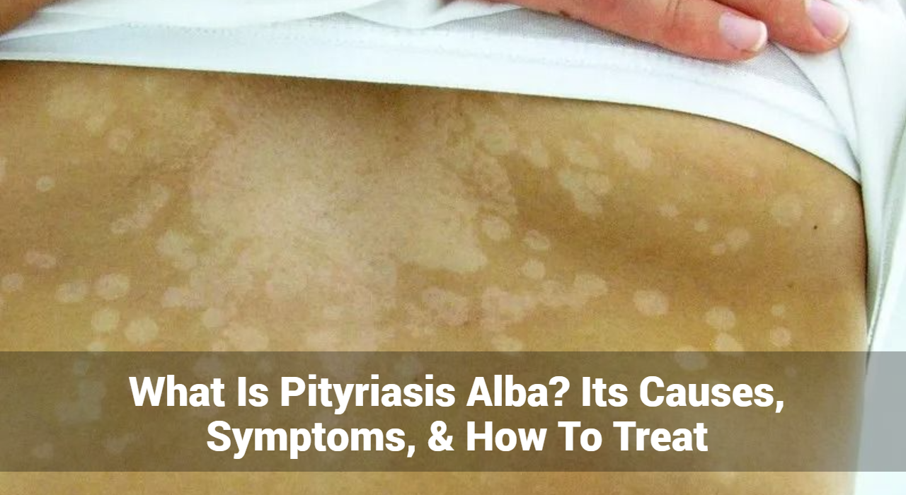
Pityriasis alba is a skin disorder that manifests as pale or light-colored patches on the skin. These patches are usually slightly scaly and have well-defined borders. The condition is most commonly observed in children and adolescents but can also affect adults. Although it often resolves on its own over time, the condition can cause cosmetic concerns and anxiety.

What Causes of Pityriasis Alba?

The exact cause of pityriasis alba is not fully understood, but several factors are believed to contribute to its development:

  1. Skin Dryness: One of the most common theories is that pityriasis alba is related to skin dryness. The patches may appear as a result of the skin’s reduced ability to retain moisture, leading to a contrast between the affected and unaffected areas.
  2. Sun Exposure: There is evidence to suggest that increased sun exposure can exacerbate or trigger pityriasis alba. The skin patches often become more noticeable after sun exposure, which can further lighten the affected areas.
  3. Eczema: Pityriasis alba may be associated with a history of eczema (atopic dermatitis). Children with eczema may develop these patches as a residual effect of their eczema.
  4. Immunological Factors: Some researchers believe that pityriasis alba may be related to an immune response that affects skin pigmentation, although more research is needed to confirm this theory.
  5. Nutritional Deficiencies: While not a primary cause, certain nutritional deficiencies, particularly in vitamins like vitamin D and calcium, may influence the appearance of pityriasis alba.

Track and Manage your Eczema treatment using a comprehensive Eczema App
Download Eczemaless now

What Are The Common Symptoms of Pityriasis Alba?

The symptoms of pityriasis alba are typically mild and may include:

  • Light-Colored Patches: The primary symptom is the presence of light-colored, slightly scaly patches on the skin. These patches are usually paler than the surrounding skin and have well-defined borders.
  • Dry, Flaky Skin: The affected skin may appear dry and flaky, although this is not always the case. The patches may be more noticeable in individuals with darker skin tones.
  • Asymptomatic: In most cases, pityriasis alba is asymptomatic, meaning it does not cause itching, pain, or other discomfort. However, some individuals may experience mild itching or irritation.
  • Common Locations: Pityriasis alba commonly appears on the face, arms, and torso. The patches may be more visible in areas that are frequently exposed to the sun.

Diagnosis of Pityriasis Alba

Diagnosing pityriasis alba typically involves a physical examination by a dermatologist. The doctor will examine the skin patches and assess their characteristics. In some cases, a skin biopsy may be performed to rule out other skin conditions that may present with similar symptoms, such as vitiligo or tinea versicolor.

Treatment Options for Pityriasis Alba

Treatment for pityriasis alba is generally not necessary, as the condition often resolves on its own over time. However, there are several treatment options available to improve the appearance of the skin and manage symptoms:

1. Moisturizers: Regular application of moisturizers can help alleviate dryness and improve the appearance of affected skin. Products containing emollients and humectants, such as ceramides or hyaluronic acid, are particularly effective in maintaining skin hydration.

2. Topical Steroids: In cases where there is significant dryness or irritation, topical corticosteroids may be prescribed. These medications help reduce inflammation and improve the appearance of the skin. Low-potency corticosteroids are typically used for this purpose.

3. Sunscreen: Using sunscreen is crucial for managing pityriasis alba. Sunscreen helps protect the skin from further sun damage and prevents the patches from becoming more noticeable. Broad-spectrum sunscreens with an SPF of 30 or higher are recommended.

4. Topical Calcineurin Inhibitors: For individuals with persistent symptoms, topical calcineurin inhibitors such as tacrolimus or pimecrolimus may be used. These medications help reduce inflammation without the side effects associated with steroids.

5. Pigment Restoring Treatments: Although not always necessary, some people seek treatments to restore skin pigmentation. Options include topical treatments containing ingredients like hydroquinone or laser therapy. These treatments should be discussed with a dermatologist to determine their suitability.


GET IN CONTROL OF YOUR ECZEMA

Use our AI tool to check the severity of Eczema and keep track of your Eczema progress.

Use our AI tool to check the severity of Eczema and keep track of your Eczema progress.


Natural Remedies for Pityriasis Alba

In addition to conventional treatments, several natural remedies may help improve the appearance of pityriasis alba and support skin health:

1. Aloe Vera: Aloe vera has moisturizing and soothing properties that can benefit dry, flaky skin. Applying pure aloe vera gel to the affected areas may help improve hydration and reduce flakiness.

2. Coconut Oil: Coconut oil is known for its moisturizing and anti-inflammatory properties. Applying coconut oil to the skin can help alleviate dryness and improve skin texture.

3. Vitamin E: Vitamin E is a powerful antioxidant that supports skin health. Applying vitamin E oil or using skincare products containing vitamin E may help improve the appearance of the skin and protect it from damage.

4. Oatmeal Baths: Oatmeal has soothing properties that can help relieve dry and itchy skin. Taking an oatmeal bath or using colloidal oatmeal in a bath can provide relief and improve skin condition.

5. Proper Hydration: Drinking plenty of water and maintaining overall hydration is essential for skin health. Proper hydration helps keep the skin moisturized from within and may contribute to the overall improvement of skin appearance.

Lifestyle Tips for Managing Pityriasis Alba

In addition to treatments and remedies, adopting certain lifestyle changes can help manage and prevent pityriasis alba:

  1. Maintain Skin Hydration: Regularly apply moisturizers to keep the skin hydrated and prevent dryness.
  2. Protect from Sun Exposure: Use sunscreen and protective clothing to shield the skin from harmful UV rays, which can exacerbate the condition.
  3. Avoid Irritants: Be mindful of skincare products that may irritate the skin. Opt for gentle, non-irritating products suitable for sensitive skin.
  4. Healthy Diet: Maintain a balanced diet rich in vitamins and minerals to support overall skin health. Foods high in antioxidants and essential fatty acids can be beneficial.
  5. Regular Skin Care: Follow a consistent skincare routine that includes cleansing and moisturizing to keep the skin healthy and resilient.

Prognosis and Outlook

Pityriasis alba is a benign condition with a generally favorable prognosis. The light-colored patches often resolve on their own over time, although the process may take several months to years. In many cases, the skin returns to its normal pigmentation without the need for medical intervention.

Conclusion

Pityriasis alba is a common skin condition characterized by light-colored patches, often affecting children and adolescents. While it is benign and typically resolves on its own, understanding its causes, symptoms, and treatment options can help manage the condition more effectively. Regular moisturization, sun protection, and appropriate treatments can improve skin appearance and alleviate any associated discomfort. If you or someone you know is experiencing symptoms of pityriasis alba, consult a dermatologist OR healthcare professional can provide personalized advice and treatment options to ensure optimal skin health and management.


Track and Manage your Eczema treatment using a comprehensive Eczema App
Download Eczemaless now

Managing Wound Infections – Symptoms, Causes, & How To Treat

Wound infections are a common medical concern that can affect anyone with a cut, scrape, or surgical incision. These infections can lead to serious complications if not properly managed, making it crucial to understand their symptoms, causes, and treatment options. In this comprehensive guide, we will explore every aspect of wound infections to help you recognize, prevent, and treat them effectively.

What Are Wound Infections?

A wound infection occurs when harmful bacteria or other pathogens invade a break in the skin, leading to an inflammatory response. The skin acts as a barrier to protect the body from external threats, and when it is compromised, it can allow microorganisms to enter and cause an infection.

Different Types of Wound Infections

  1. Superficial Infections: These infections affect only the outer layers of the skin. They are usually less severe and include conditions like impetigo or cellulitis.
  2. Deep Infections: These involve deeper tissues, including muscles and bones. They can be more serious and include conditions such as osteomyelitis or septic arthritis.
  3. Surgical Site Infections (SSIs): These occur after surgical procedures and can affect the incision area. They are categorized into three types: superficial, deep, and organ/space infections.

Track and Manage your Eczema treatment using a comprehensive Eczema App
Download Eczemaless now

What Are The Symptoms of Wound Infections?

Identifying the symptoms of a wound infection early is crucial for prompt and effective treatment. Wound infections can range from mild to severe and may present with various signs. Here’s an in-depth look at the symptoms you should be aware of:

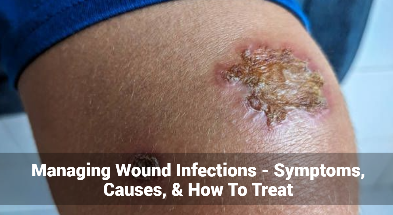
  • Redness: Redness around the wound is a common early sign of infection. This occurs due to increased blood flow to the area as part of the body’s inflammatory response. If the redness expands beyond the immediate area of the wound, it may indicate that the infection is spreading.
  • Swelling: Swelling, or edema, is another key symptom. Inflammation from the infection causes the tissues around the wound to swell. This can make the wound appear larger than initially and may affect nearby areas.
  • Heat: The affected area may feel warm or hot to the touch. This increase in temperature is a result of increased blood flow and the body’s response to the infection.
  • Pain: Pain or tenderness around the wound is common. The level of pain can vary from mild to severe and is often an indicator of inflammation and irritation in the infected area.
  • Pus or Discharge: One of the most telling signs of infection is the presence of pus or other abnormal discharge. Pus is a thick, yellowish fluid that consists of dead cells, bacteria, and tissue debris. Discharge may also be greenish or cloudy and can have a foul odor.
  • Fever: Infections can lead to systemic symptoms like fever. A rise in body temperature can be a sign that the infection is affecting the whole body. Fever often accompanies other symptoms like chills and general malaise.
  • Increased Wound Size: An infected wound might start to grow or fail to heal as expected. If the wound seems to be expanding or if the edges look swollen and irregular, it may be a sign of infection.
  • Delayed Healing: Wounds that do not show signs of improvement or continue to deteriorate despite proper care might be infected. Healing delays can be an indication that the infection is preventing the wound from closing properly.
  • Discolored Skin: The skin around the wound might change color, appearing darker or purplish. This can indicate a more severe infection or compromised blood flow to the area.
  • General Malaise: In some cases, an infection can cause a general feeling of illness or unwellness. This can include symptoms like fatigue, weakness, and a lack of appetite.

What Causes of Wound Infections?

Several factors can contribute to the development of a wound infection:

  1. Bacteria: The most common pathogens include Staphylococcus aureus, including methicillin-resistant Staphylococcus aureus (MRSA), and Streptococcus species. These bacteria are often found on the skin and can invade through breaks in the skin.
  2. Fungi: In some cases, fungi like Candida can cause infections, particularly in people with weakened immune systems.
  3. Viruses: Although less common, viral infections can occur, especially in immunocompromised individuals.
  4. Poor Wound Care: Inadequate cleaning, improper dressing, and failure to monitor the wound can increase the risk of infection.
  5. Underlying Health Conditions: Diabetes, vascular disease, and immune system disorders can impair the body’s ability to heal and fight infections.
  6. Foreign Bodies: Objects such as splinters or surgical implants left in the wound can harbor bacteria and lead to infection.


GET IN CONTROL OF YOUR ECZEMA

Use our AI tool to check the severity of Eczema and keep track of your Eczema progress.

Use our AI tool to check the severity of Eczema and keep track of your Eczema progress.


What Are The Treatment Options of Wound Infections?

The treatment of wound infections depends on the severity and type of infection. Here are common approaches:

1. Cleaning the Wound

  • Initial Cleaning: Use clean water or saline solution to gently clean the wound. Avoid using hydrogen peroxide or iodine, as these can damage healthy tissue.
  • Debridement: In some cases, removal of dead tissue or foreign bodies may be necessary.

2. Antibiotic Therapy

  • Topical Antibiotics: For superficial infections, topical antibiotics like mupirocin or bacitracin may be prescribed.
  • Oral Antibiotics: More severe or deep infections may require oral antibiotics. Your healthcare provider will choose an antibiotic based on the specific bacteria involved.

3. Surgical Intervention

  • Incision and Drainage: If an abscess forms, a procedure to drain the pus may be required.
  • Surgical Debridement: For deep infections or necrotizing fasciitis, surgical removal of infected tissue may be necessary.

4. Wound Care

  • Dressing Changes: Regularly change the wound dressing to keep the wound clean and dry.
  • Monitoring: Keep an eye on the wound for signs of worsening infection or complications.

5. Pain Management

  • Analgesics: Over-the-counter pain relievers like ibuprofen or acetaminophen can help manage pain and inflammation.

6. Managing Underlying Conditions

  • Diabetes Management: Control blood sugar levels to aid in wound healing.
  • Improving Circulation: Address vascular issues to promote healing.

How Can You Prevent From Wound Infections?

Preventing wound infections is essential, especially for those with chronic conditions or those undergoing surgery. Here are some tips:

  • Proper Wound Care: Clean wounds immediately and apply antiseptic if recommended. Keep the wound covered with a sterile bandage.
  • Hygiene: Practice good hand hygiene to avoid transferring bacteria to wounds.
  • Healthy Lifestyle: Maintain a balanced diet, stay hydrated, and avoid smoking to support overall health and immune function.
  • Monitor Chronic Conditions: Manage chronic diseases like diabetes to prevent complications.

When to Seek Medical Attention

Not all wounds will develop infections, but it’s important to seek medical help if you experience any of the following:

  • Persistent Symptoms: Symptoms of infection that do not improve with basic care.
  • Severe Pain: Intense pain that worsens over time.
  • Fever: A high fever or chills along with the wound infection.
  • Rapidly Worsening Condition: Signs that the infection is spreading or worsening.

Conclusion

Wound infections, while common, require careful attention to avoid serious complications. By understanding the symptoms, causes, and treatments, you can take proactive steps to manage and prevent these infections. Always consult a dermatologist or medical advice if you have concerns about a wound infection, especially if symptoms are severe or persistent. Proper wound care, timely treatment, and attention to underlying health conditions are key to a successful recovery.


Track and Manage your Eczema treatment using a comprehensive Eczema App
Download Eczemaless now

What Is Eczema Herpeticum? It’s Symptoms, Causes, and treatment

Eczema Herpeticum is a potentially serious condition that arises when the herpes simplex virus (HSV) infects skin that is already affected by eczema. This article delves into the intricacies of Eczema Herpeticum, covering its symptoms, causes, and treatment options.

What is Eczema Herpeticum?

Eczema Herpeticum occurs when herpes simplex virus, commonly known for causing cold sores or genital herpes, infects areas of skin that are compromised due to eczema. Eczema, also known as atopic dermatitis, is a chronic condition characterized by inflamed, itchy, and often cracked skin. When this compromised skin comes into contact with the herpes virus, it can lead to a severe and sometimes life-threatening infection.

What Are The Symptoms of Eczema Herpeticum?

The symptoms of Eczema Herpeticum can vary but generally include:

  1. Fever and Malaise: Often, the first sign of Eczema Herpeticum is a fever, particularly in young children. This is accompanied by general malaise and fatigue.
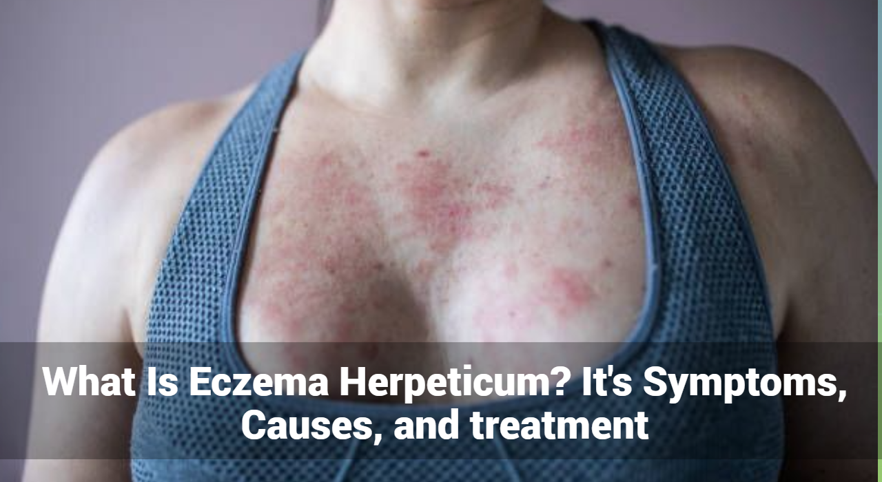
  2. Vesicular Lesions: The hallmark of Eczema Herpeticum is the appearance of small, fluid-filled blisters or vesicles. These blisters are often clustered and can be found on the skin affected by eczema.
  3. Redness and Inflammation: The areas around the vesicles are usually red and inflamed. The infection can exacerbate existing eczema, leading to increased redness and swelling.
  4. Pain and Itching: The affected skin can become very painful and itchy. The discomfort is typically more intense than that caused by eczema alone.
  5. Crusting and Weeping: As the vesicles break, they can form crusts and weep, which can increase the risk of secondary bacterial infections.
  6. Enlarged Lymph Nodes: In some cases, the lymph nodes near the affected area may become swollen as the body responds to the infection.
  7. Systemic Symptoms: In severe cases, symptoms might include systemic issues such as fever, chills, and headache, indicating that the virus might be spreading beyond the skin.

Track and Manage your Eczema treatment using a comprehensive Eczema App
Download Eczemaless now

What Causes of Eczema Herpeticum?

The primary cause of Eczema Herpeticum is infection with the herpes simplex virus (HSV). HSV is categorized into two types:

  1. HSV Type 1: Often associated with oral herpes, this type is the most common cause of Eczema Herpeticum.
  2. HSV Type 2: Primarily linked with genital herpes, it can also cause Eczema Herpeticum, though this is less common.

People with eczema have skin that is more vulnerable to infections due to its compromised barrier function. When the herpes virus comes into contact with eczema-affected skin, it can rapidly proliferate, leading to the development of Eczema Herpeticum.

Risk Factors

Several factors increase the risk of developing Eczema Herpeticum:

  1. Compromised Immune System: Individuals with weakened immune systems are at higher risk for severe infections, including Eczema Herpeticum.
  2. Active Eczema: Areas of active eczema are particularly susceptible to viral infections.
  3. Close Contact with Infected Individuals: Herpes simplex is highly contagious and can spread through close personal contact with someone who has an active outbreak.
  4. Young Age: Children, particularly those under the age of five, are at higher risk. Their immune systems are still developing, making them more vulnerable to severe infections.
  5. Recent Herpes Exposure: Individuals who have recently been exposed to herpes simplex virus or who have a history of herpes infections are at increased risk.

Diagnosis of Eczema Herpeticum

Diagnosing Eczema Herpeticum typically involves:

  1. Clinical Examination: A healthcare provider will examine the appearance of the skin and the characteristic symptoms, such as the presence of vesicular lesions.
  2. Laboratory Tests: To confirm the diagnosis, a sample may be taken from the lesions for laboratory testing. This can include:
    • PCR Testing: Polymerase Chain Reaction (PCR) tests can detect herpes simplex DNA.
    • Viral Culture: A culture of the fluid from the blisters can help identify the virus.
    • Direct Fluorescent Antibody (DFA) Testing: This test can identify the presence of HSV antigens.

GET IN CONTROL OF YOUR ECZEMA

Use our AI tool to check the severity of Eczema and keep track of your Eczema progress.

Use our AI tool to check the severity of Eczema and keep track of your Eczema progress.


How Can Treatment of Eczema Herpeticum?

Immediate medical attention is crucial in managing Eczema Herpeticum to prevent complications. Treatment usually involves:

  1. Antiviral Medications: Oral antiviral drugs such as acyclovir, valacyclovir, or famciclovir are commonly prescribed. These medications help to reduce the severity and duration of the infection.
  2. Topical Antivirals: In some cases, topical antiviral creams may be applied to the lesions to alleviate symptoms and prevent the spread of the virus.
  3. Supportive Care: Symptomatic relief can be provided with medications to manage pain, itching, and fever. Over-the-counter pain relievers such as ibuprofen or acetaminophen can be helpful.
  4. Skin Care: Keeping the affected skin clean and dry is important. Applying moisturizers and avoiding irritants can help manage eczema symptoms and prevent further infection.
  5. Preventive Measures: Avoiding close contact with others while lesions are present can prevent the spread of HSV. Good hygiene practices, such as regular hand washing and avoiding touching the face, can also reduce the risk of transmission.
  6. Monitoring and Follow-Up: Regular follow-up with a healthcare provider is important to monitor the progress of the infection and to adjust treatment as needed.

Prevention of Eczema Herpeticum

Preventing Eczema Herpeticum involves:

  1. Managing Eczema: Effective management of eczema can help reduce the risk of developing Eczema Herpeticum. This includes using appropriate eczema treatments and avoiding known triggers.
  2. Avoiding Herpes Exposure: Minimizing contact with individuals who have active herpes infections can reduce the risk of exposure. Individuals with a history of herpes simplex should be particularly cautious.
  3. Hygiene Practices: Practicing good hygiene, including frequent hand washing and avoiding touching the face, can help prevent the spread of the virus.
  4. Education: Educating those with eczema and their caregivers about the signs and symptoms of herpes simplex infection can facilitate early detection and treatment.

Conclusion

Eczema Herpeticum is a serious condition that requires prompt medical attention. Understanding the symptoms, causes, and treatment options is crucial for effective management. By following preventive measures and seeking timely treatment, individuals with eczema can reduce their risk of developing this potentially severe infection and maintain better overall skin health. If you suspect you or someone you know may have Eczema Herpeticum, it is essential to consult a dermatologist for appropriate diagnosis and treatment.


Track and Manage your Eczema treatment using a comprehensive Eczema App
Download Eczemaless now

Kawasaki Disease in Children | Symptoms, Causes, Treatment

Kawasaki Disease, also known as Kawasaki Syndrome, is a rare but serious condition that primarily affects children. It is characterized by inflammation of the blood vessels throughout the body, leading to a range of symptoms that can have significant long-term effects if not addressed promptly. Understanding the symptoms, causes, and treatment options for Kawasaki Disease is crucial for parents, caregivers, and healthcare professionals alike.

What is Kawasaki Disease?

Kawasaki Disease is an autoimmune skin condition that causes inflammation in the walls of medium-sized arteries, including those in the heart. It primarily affects children under the age of five and can lead to serious complications if not treated promptly. The disease was first identified by Dr. Tomisaku Kawasaki in Japan in the 1960s, and it has since been recognized globally.

What Are Symptoms of Kawasaki Disease?

The symptoms of Kawasaki Disease typically occur in phases and can vary from child to child. Early recognition and treatment are crucial to prevent complications. Here are the common symptoms associated with Kawasaki Disease:

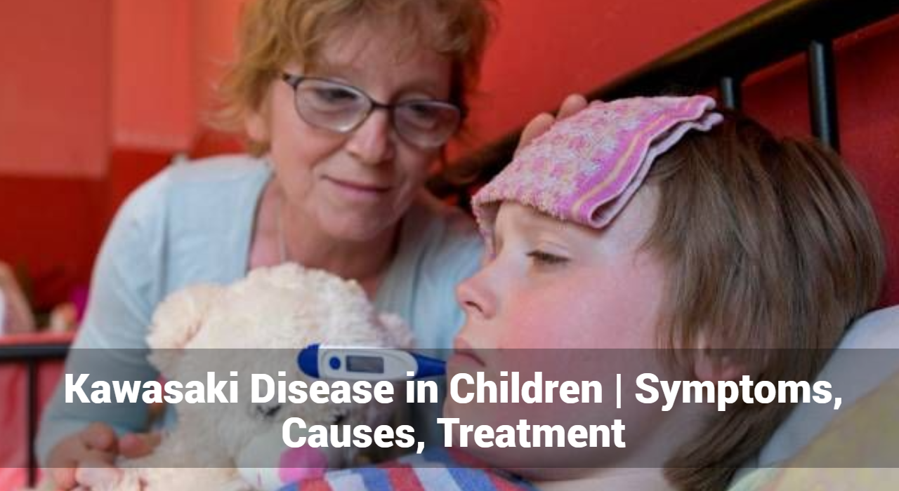
1. Fever: One of the earliest signs of Kawasaki Disease is a high fever lasting more than five days, often reaching temperatures of 102°F (38.9°C) or higher. The fever may come and go, and it is usually unresponsive to standard antipyretic medications.

2. Rash: Children with Kawasaki Disease often develop a rash that may cover large areas of the body. The rash can be red, bumpy, and may appear on the trunk, arms, and legs. It often resembles a measles-like rash and can vary in appearance.

3. Swollen Hands and Feet: Swelling of the hands and feet is another common symptom. The skin may appear red and swollen, and the child may experience discomfort and difficulty moving their limbs.

4. Conjunctivitis: Conjunctivitis, or redness and inflammation of the eyes, is frequently observed in Kawasaki Disease. The eyes may appear red and watery, but there is usually no discharge or crusting.

5. Strawberry Tongue: A distinctive symptom of Kawasaki Disease is a “strawberry tongue,” characterized by a bright red, swollen tongue with prominent taste buds. This appearance is often coupled with cracked and dry lips.

6. Swollen Lymph Nodes: Swelling of the lymph nodes, particularly those in the neck, can occur. This swelling is usually painless but can be noticeable and tender.

7. Irritability and General Malaise: Children with Kawasaki Disease may exhibit irritability and general discomfort. They may also experience joint pain and stomach pain, contributing to their overall malaise.


Track and Manage your Eczema treatment using a comprehensive Eczema App
Download Eczemaless now

What Causes of Kawasaki Disease?

The exact cause remains unknown, but several factors are believed to contribute to its development:

1. Genetic Factors: Genetics may play a role, as it tends to occur more frequently in certain populations. Children of Asian descent, particularly those of Japanese or Korean heritage, have a higher incidence of the disease.

2. Infections: Some researchers suggest that infections, such as viral or bacterial infections may trigger Kawasaki Disease. However, no specific pathogen has been conclusively linked to the disease.

3. Immune System Response: Kawasaki Disease is considered an autoimmune condition, where the body’s immune system mistakenly attacks its own blood vessels. This abnormal immune response leads to inflammation and damage to the arteries.

4. Environmental Factors: Environmental factors, such as exposure to pollutants or certain chemicals, may also play a role in the development of Kawasaki Disease. However, these factors have not been definitively proven to cause the condition.

Diagnosis of Kawasaki Disease

Diagnosing Kawasaki Disease can be challenging due to its overlapping symptoms with other illnesses. Healthcare professionals use a combination of clinical criteria and diagnostic tests to confirm the diagnosis:

1. Clinical Criteria: Diagnosis is primarily based on clinical criteria, including the presence of fever lasting more than five days along with at least four of the following symptoms:

  • Rash
  • Swelling of hands and feet
  • Conjunctivitis
  • Strawberry tongue
  • Swollen lymph nodes

2. Laboratory Tests: Blood tests may be conducted to assess inflammation markers, such as erythrocyte sedimentation rate (ESR) or C-reactive protein (CRP). Elevated levels of these markers can support the diagnosis of Kawasaki Disease.

3. Imaging Studies: Echocardiography (ultrasound of the heart) is used to evaluate any potential damage to the heart or coronary arteries. This helps in assessing the risk of complications such as coronary artery aneurysms.

GET IN CONTROL OF YOUR ECZEMA

Use our AI tool to check the severity of Eczema and keep track of your Eczema progress.

Use our AI tool to check the severity of Eczema and keep track of your Eczema progress.


What Are The Treatment of Kawasaki Disease?

Early treatment is crucial to reduce the risk of complications and improve outcomes. The primary goals of treatment are to reduce inflammation, prevent damage to the heart, and alleviate symptoms. Treatment options include:

1. Intravenous Immunoglobulin (IVIG): Intravenous immunoglobulin (IVIG) is the primary treatment for Kawasaki Disease. It is administered through an intravenous drip and helps reduce inflammation, lower fever, and prevent coronary artery damage. The treatment is most effective when given within the first ten days of the illness.

2. Aspirin: Aspirin is used in conjunction with IVIG to help reduce inflammation and fever. It also plays a role in preventing blood clot formation and reducing the risk of coronary artery complications. The dosage and duration of aspirin therapy are carefully managed by healthcare professionals.

3. Steroids: In some cases, corticosteroids may be prescribed if the initial treatment with IVIG and aspirin is not effective. Steroids help control inflammation and can be beneficial in severe cases of Kawasaki Disease.

4. Supportive Care: Supportive care includes managing symptoms and providing comfort measures. This may involve hydration, pain management, and monitoring for any potential side effects of treatment.

5. Long-Term Follow-Up: Regular follow-up appointments are essential to monitor the child’s heart health and ensure there are no long-term complications. This may include periodic echocardiograms to assess the condition of the coronary arteries.

Complications of Kawasaki Disease

Without timely and appropriate treatment, Kawasaki Disease can lead to several serious complications:

1. Coronary Artery Aneurysms: The most significant complication of Kawasaki Disease is the formation of aneurysms in the coronary arteries. These aneurysms can lead to long-term heart problems, including heart attacks and reduced heart function.

2. Myocarditis: Inflammation of the heart muscle (myocarditis) can occur, leading to weakened heart function and potential heart failure.

3. Heart Valve Issues: In some cases, Kawasaki Disease can affect the heart valves, leading to valvular dysfunction or regurgitation.

4. Long-Term Cardiovascular Risk: Children who have had Kawasaki Disease may be at increased risk for cardiovascular issues later in life. Ongoing monitoring and management are essential to mitigate these risks.

Conclusion

Kawasaki Disease is a serious condition that requires prompt recognition and treatment to prevent complications. Understanding the symptoms, causes, and available treatments can help ensure that affected children receive the care they need. If you suspect your child may have to seek medical attention immediately to receive a timely diagnosis and appropriate treatment. With early intervention, many children recover fully and lead healthy lives.

For more information, consult a dermatologist to stay informed about the latest research and treatment options.


Track and Manage your Eczema treatment using a comprehensive Eczema App
Download Eczemaless now


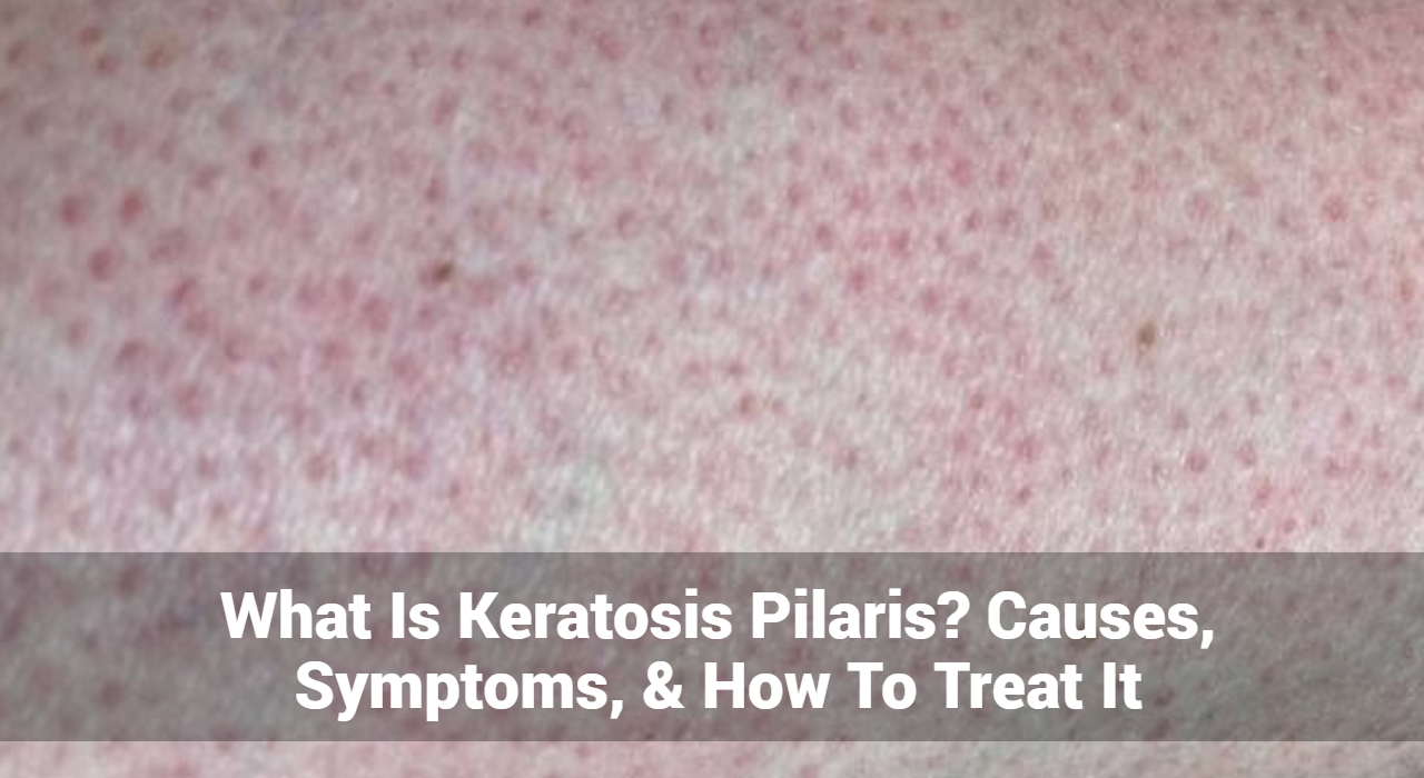
What Is Keratosis Pilaris? Causes, Symptoms, & Treatment

Keratosis pilaris (KP), often referred to as “chicken skin,” is a common and benign skin condition that manifests as small, rough bumps on the skin. Despite its prevalence, it is frequently misunderstood, and its impact on those affected can range from purely cosmetic to a source of discomfort. This comprehensive guide explores keratosis pilaris in detail, including its causes, symptoms, and treatment options.

What Is Keratosis Pilaris?

Keratosis pilaris is a skin condition characterized by the appearance of small, gooseflesh-like bumps that often give the skin a rough texture. These bumps are caused by the accumulation of keratin, a protein found in the skin, in hair follicles. The condition is typically harmless and not associated with any underlying health problems, but it can be cosmetically concerning for many people.

KP commonly affects areas where hair follicles are present, such as the upper arms, thighs, buttocks, and sometimes the face. It is not contagious and generally does not pose any health risks.

What Causes of Keratosis Pilaris?

The exact cause of keratosis pilaris is not fully understood, but several factors are believed to contribute to its development:

  1. Hereditary Factors: KP often runs in families, suggesting a genetic predisposition. If your parents or siblings have KP, you might be more likely to develop it.
  2. Excess Keratin Production: The primary cause of KP is the overproduction of keratin, which clogs the hair follicles. This buildup forms the characteristic bumps on the skin.
  3. Associated Conditions: KP is often seen in conjunction with other skin conditions, such as eczema or dry skin. People with these conditions may be more prone to developing KP.
  4. Hormonal Influences: Changes in hormone levels, particularly during puberty or pregnancy, may exacerbate the condition, although KP can occur at any age.
  5. Dry Skin: Cold weather and low humidity can lead to dry skin, which may worsen the appearance of KP. Conversely, warmer weather often improves skin texture.

Track and Manage your Eczema treatment using a comprehensive Eczema App
Download Eczemaless now


What Are The Symptoms of Keratosis Pilaris?

Keratosis pilaris typically presents with specific symptoms that make it identifiable:

  1. Appearance: The most common symptom is the presence of small, round bumps that can be white, red, or skin-colored. These bumps are usually found on the upper arms, thighs, buttocks, and sometimes the face.
  2. Skin Feel: The affected areas of the skin often feel rough or gritty to the touch, similar to sandpaper.
  3. Accompanying Sensations: The bumps may be accompanied by dry or itchy skin, although KP itself is usually not painful.
  4. Aesthetic Impact: While KP is harmless, many individuals seek treatment due to its cosmetic appearance, which can affect self-esteem and confidence.
  5. Harmless Nature: KP does not cause pain, swelling, or significant discomfort, although the cosmetic impact may be distressing for some.

What Are The Treatment Options for Keratosis Pilaris?

While keratosis pilaris cannot be cured, various treatments can help manage and improve the appearance of the skin. Treatment options include:

Topical Treatments

  • Exfoliating Agents: Products containing exfoliating ingredients can help remove dead skin cells and reduce the buildup of keratin. Commonly used exfoliants include:
  1. Salicylic Acid: Helps to break down keratin plugs and reduce inflammation.
  2. Alpha Hydroxy Acids (AHAs): Lactic acid or glycolic acid can gently exfoliate the skin and improve texture.
  3. Urea: Hydrates and softens the skin, making it easier to slough off dead cells.
  • Retinoids: Topical retinoids, such as tretinoin, can help unclog hair follicles and improve skin cell turnover. These are available by prescription and should be used under a dermatologist’s guidance.

Moisturizers

  • Hydrating Creams: Regular use of moisturizers can help alleviate dryness and improve the appearance of KP. Look for products with ingredients like:
  1. Ceramides: Help restore the skin barrier and retain moisture.
  2. Hyaluronic Acid: Provides deep hydration and improves skin texture.
  3. Glycolic Acid: A mild exfoliant that also acts as a moisturizer.

Laser Therapy

  • Targeted Treatments: Laser treatments can be used to reduce redness and improve skin texture by targeting and destroying the excess keratin and inflammation. This option is usually considered for more severe cases or when other treatments have not been effective.

Chemical Peels

  • Professional Peels: Chemical peels performed by dermatologists can help exfoliate the top layer of skin, improving texture and appearance. Peels containing salicylic acid, glycolic acid, or lactic acid can be beneficial.

Lifestyle Adjustments

  • Regular Exfoliation: Incorporating gentle exfoliation into your skincare routine can help manage KP. Avoid harsh scrubbing, which can irritate the skin.
  • Hydration: Maintaining adequate hydration by drinking plenty of water and using moisturizing products helps keep the skin healthy and less prone to dryness.

Home Remedies

  • Natural Exfoliants: Some people find relief with natural exfoliating remedies, such as sugar scrubs or oatmeal baths. However, these should be used cautiously to avoid skin irritation.

GET IN CONTROL OF YOUR ECZEMA

Use our AI tool to check the severity of Eczema and keep track of your Eczema progress.

Use our AI tool to check the severity of Eczema and keep track of your Eczema progress.


Diagnosis of Keratosis Pilaris

Diagnosing keratosis pilaris is typically straightforward, based on visual examination and patient history. Consult a dermatologist may use the following methods to confirm the diagnosis:

  • Physical Inspection: The dermatologist will inspect the skin for the characteristic bumps and rough texture associated with KP.
  • Patient Background: Discussion of symptoms, family history, and any related skin conditions helps in making an accurate diagnosis.
  • Ruling Out Other Conditions: In some cases, a dermatologist may need to differentiate KP from other skin conditions that present with similar symptoms, such as acne or folliculitis.
  • Biopsy: A skin biopsy is rarely necessary but may be performed if the diagnosis is uncertain or if there are unusual symptoms.

Preventing Keratosis Pilaris

While keratosis pilaris cannot be completely prevented, certain measures can help minimize its appearance and reduce flare-ups:

  • Hydration: Keep your skin well-moisturized to prevent dryness, which can exacerbate KP. Use thick, hydrating creams or lotions, especially during colder months.
  • Gentle Products: Use mild, non-irritating skincare products that do not strip the skin of its natural oils.
  • Avoid Over-Exfoliation: Exfoliate the skin gently to prevent irritation. Over-exfoliating can lead to further inflammation and worsening of KP.
  • Sunblock: Protect your skin from excessive sun exposure, which can worsen skin dryness and irritation.
  • Associated Skin Conditions: If you have other skin conditions like eczema or psoriasis, managing these effectively can help reduce the impact of KP.

Conclusion

Keratosis pilaris is a common and generally harmless skin condition characterized by small, rough bumps caused by the buildup of keratin in hair follicles. While it cannot be cured, effective management through topical treatments, moisturizers, and lifestyle adjustments can significantly improve skin appearance and texture. Consulting a dermatologist can provide personalized treatment options and guidance, especially for more severe cases or when standard treatments are ineffective. By understanding the causes, symptoms, and treatment options, individuals with KP can take proactive steps to manage their condition and enhance their skin health.


Track and Manage your Eczema treatment using a comprehensive Eczema App
Download Eczemaless now