What Is Venous Disease? Its Types, Causes, Symptoms & Treatment

Venous disease, also known as venous insufficiency, is a condition in which the veins in the body do not properly return blood to the heart. This can lead to a range of symptoms and complications, from mild discomfort to severe health issues. Understanding the types, causes, symptoms, and treatment options for venous disease is crucial for effective management and prevention.

What is Venous Disease?

Venous disease occurs when veins become damaged or dysfunctional, affecting their ability to transport blood efficiently. This condition can affect any veins in the body, but it most commonly impacts those in the legs. Veins have one-way valves that prevent blood from flowing backward. When these valves fail, blood can pool in the veins, leading to various complications.

Common Types of Venous Disease

  1. Varicose Veins
    • Description: Enlarged, twisted veins that are often visible under the skin.
    • Symptoms: Aching, swelling, and a feeling of heaviness in the legs. They may also cause itching and skin discoloration.
  2. Spider Veins
    • Description: Small, red, blue, or purple veins that appear close to the surface of the skin.
    • Symptoms: Typically asymptomatic but can cause cosmetic concerns and mild discomfort.
  3. Chronic Venous Insufficiency (CVI)
    • Description: A long-term condition where the veins cannot pump enough blood back to the heart.
    • Symptoms: Swelling, skin changes, and ulcers on the legs.
  4. Deep Vein Thrombosis (DVT)
    • Description: A blood clot that forms in a deep vein, usually in the legs.
    • Symptoms: Swelling, pain, and redness in the affected leg. DVT can lead to serious complications if the clot travels to the lungs (pulmonary embolism).
  5. Superficial Thrombophlebitis
    • Description: Inflammation and clotting in a superficial vein.
    • Symptoms: Redness, swelling, and pain along the affected vein.

What Causes of Venous Disease?

01. Genetics

  • A family history of venous disease increases the risk of developing the condition.

02. Age

  • The risk of venous disease increases with age due to the weakening of vein walls and valves over time.

03. Gender

  • Women are more likely to develop venous disease, partly due to hormonal changes related to pregnancy, menopause, and the use of birth control pills.

04. Prolonged Standing or Sitting

  • Jobs that require long periods of standing or sitting can contribute to venous disease by hindering blood flow.

05. Obesity

  • Excess weight puts additional pressure on veins, increasing the risk of venous disease.

06. Pregnancy

  • Pregnancy increases blood volume and puts additional pressure on veins, leading to an increased risk of venous disease.

07. Injury or Surgery

  • Damage to veins from injury or surgery can lead to venous insufficiency.

08. Lack of Exercise

  • Regular exercise helps maintain healthy blood flow. A sedentary lifestyle can contribute to venous disease.

Track and Manage your Eczema treatment using a comprehensive Eczema App
Download Eczemaless now


What Are The Symptoms of Venous Disease?

1. Swelling

  • Typically in the legs and ankles, swelling is a common symptom of venous disease.

2. Pain and Discomfort

  • Aching, throbbing, or a feeling of heaviness in the legs.

3. Skin Changes

  • Discoloration, dryness, or the development of ulcers, particularly around the ankles.

4. Visible Veins

  • Enlarged, twisted, or bulging veins (varicose veins) and small, web-like veins (spider veins).

5. Itching and Burning

  • A sensation of itching or burning in the affected areas.

6. Leg Cramps

  • Particularly at night, leg cramps can be a symptom of venous disease.

7. Restless Legs

  • A compelling urge to move the legs, often accompanied by uncomfortable sensations.

Diagnosis of Venous Disease

Diagnosing venous disease typically involves a physical examination and medical history review. Additional diagnostic tests may include:

  1. Duplex Ultrasound
    • This non-invasive test uses sound waves to visualize blood flow in the veins and detect clots or blockages.
  2. Venography
    • A special dye is injected into the veins, and X-rays are taken to visualize the veins and identify any abnormalities.
  3. Magnetic Resonance Venography (MRV)
    • An MRI scan that provides detailed images of the veins.

What Are The Treatment Options for Venous Disease?

1. Lifestyle Changes

  • Exercise: Regular physical activity helps improve blood flow and strengthen the calf muscles, which play a crucial role in venous return.
  • Diet: Maintaining a healthy weight and eating a balanced diet can reduce pressure on the veins.
  • Elevating the Legs: Elevating the legs above the heart level can help reduce swelling and improve blood flow.

2. Compression Therapy

  • Compression Stockings: Specially designed stockings apply pressure to the legs, helping veins move blood more efficiently. They come in different levels of compression and lengths.

3. Medications

  • Blood Thinners: Used to treat and prevent blood clots in conditions like DVT.
  • Anti-inflammatory Medications: Help reduce pain and swelling associated with venous disease.

4. Minimally Invasive Procedures

  • Sclerotherapy: A solution is injected into the affected veins, causing them to collapse and eventually be absorbed by the body.
  • Endovenous Laser Treatment (EVLT): A laser fiber is inserted into the vein, emitting energy that causes the vein to collapse.
  • Radiofrequency Ablation (RFA): Similar to EVLT, this procedure uses radiofrequency energy to heat and close off the vein.

5. Surgical Treatments

  • Vein Stripping: Removal of the affected vein through small incisions.
  • Ambulatory Phlebectomy: Removal of varicose veins through tiny punctures in the skin.
  • Vein Bypass: Creating a bypass around the damaged vein using a healthy vein from another part of the body.

How Can Prevent of Venous Disease

1. Stay Active

  • Regular exercise, such as walking, swimming, or cycling, promotes healthy blood flow.

2. Maintain a Healthy Weight

  • Keeping a healthy weight reduces pressure on the veins.

3. Avoid Prolonged Standing or Sitting

  • Take breaks to walk around or stretch if your job requires long periods of sitting or standing.

4. Elevate Your Legs

  • Elevate your legs when resting to help reduce swelling and improve circulation.

5. Wear Compression Stockings

  • Compression stockings can help prevent venous disease, especially if you are at high risk.

6. Stay Hydrated

  • Drinking plenty of water helps maintain healthy blood circulation.

GET IN CONTROL OF YOUR ECZEMA

Use our AI tool to check the severity of Eczema and keep track of your Eczema progress.

Use our AI tool to check the severity of Eczema and keep track of your Eczema progress.


Home Remedies for Venous Disease

Venous disease, which encompasses conditions such as varicose veins, spider veins, and chronic venous insufficiency, can be managed effectively with lifestyle changes and home remedies. While medical treatments are often necessary, incorporating home remedies can provide relief from symptoms and improve overall vascular health. Here are some effective home remedies for venous disease.

1. Compression Stockings

How They Help: Compression stockings apply pressure to the legs, helping veins move blood more efficiently back to the heart. They can reduce swelling, discomfort, and the risk of developing further venous complications.

Usage:

  • Wear compression stockings as directed by your healthcare provider, usually throughout the day.
  • Ensure they fit properly to avoid discomfort or skin damage.

2. Elevate Your Legs

How It Helps: Elevating your legs above heart level helps reduce swelling and improves blood flow by using gravity to assist venous return.

Usage:

  • Lie down and prop your legs on pillows or a cushion.
  • Elevate your legs for 15-30 minutes several times a day.

3. Regular Exercise

How It Helps: Exercise promotes healthy blood circulation and strengthens the calf muscles, which play a crucial role in pushing blood back to the heart.

Suggestions:

  • Engage in low-impact activities such as walking, swimming, or cycling.
  • Aim for at least 30 minutes of exercise most days of the week.

4. Maintain a Healthy Weight

How It Helps: Excess weight puts additional pressure on veins, exacerbating venous disease symptoms.

Tips:

  • Follow a balanced diet rich in fruits, vegetables, lean proteins, and whole grains.
  • Monitor portion sizes and avoid high-calorie, low-nutrient foods.

5. Herbal Remedies

a. Horse Chestnut Extract:

  • How It Helps: Horse chestnut contains aescin, which helps improve venous tone and reduce swelling.
  • Usage: Available in supplement form or topical creams. Consult with a healthcare provider for proper dosage.

b. Grape Seed Extract:

  • How It Helps: Rich in antioxidants, grape seed extract can help strengthen blood vessels and reduce inflammation.
  • Usage: Take as a supplement, following the recommended dosage on the packaging.

c. Gotu Kola:

  • How It Helps: Gotu kola may improve circulation and reduce swelling.
  • Usage: Available as a supplement or in topical creams. Follow dosage recommendations.

6. Dietary Changes

How It Helps: A diet rich in certain nutrients can support vein health and reduce symptoms of venous disease.

Recommendations:

  • Fiber: Prevents constipation, reducing pressure on veins. Include foods like whole grains, fruits, and vegetables.
  • Flavonoids: Strengthen blood vessels and improve circulation. Found in foods like berries, citrus fruits, and dark chocolate.
  • Vitamin C: Supports collagen production, essential for vein health. Include citrus fruits, strawberries, and bell peppers.
  • Vitamin E: Improves blood flow. Found in nuts, seeds, and leafy greens.

7. Hydration

How It Helps: Staying hydrated helps maintain healthy blood circulation and prevents blood from becoming too thick.

Tips:

  • Drink plenty of water throughout the day.
  • Limit intake of dehydrating beverages like alcohol and caffeine.

8. Avoid Prolonged Sitting or Standing

How It Helps: Prolonged periods of sitting or standing can hinder blood flow and exacerbate venous disease symptoms.

Tips:

  • Take breaks to walk around or stretch every hour if you have a sedentary job.
  • Shift your weight and move your legs regularly if you must stand for long periods.

9. Wear Loose Clothing

How It Helps: Tight clothing, especially around the waist and legs, can restrict blood flow and worsen venous disease.

Tips:

  • Opt for loose-fitting, comfortable clothing.
  • Avoid tight belts, leggings, or stockings that aren’t designed for compression therapy.

10. Massage

How It Helps: Gentle massage can help improve circulation and reduce swelling in the legs.

Tips:

  • Use upward strokes, starting from the ankles and moving towards the heart.
  • Use a moisturizing lotion or oil to reduce friction and improve comfort.

11. Essential Oils

How They Help: Certain essential oils can promote circulation and reduce swelling.

Suggestions:

  • Cypress Oil: Known for its circulation-boosting properties. Dilute with a carrier oil and massage into the affected area.
  • Lavender Oil: Provides relief from pain and inflammation. Dilute and apply as needed.
  • Peppermint Oil: Offers a cooling effect and helps reduce swelling. Dilute before use.

12. Hydrotherapy

How It Helps: Alternating between hot and cold water can improve circulation and reduce swelling.

Usage:

  • End your shower with a blast of cold water on your legs.
  • Use warm water for a few minutes, followed by cold water for a minute. Repeat this cycle several times.

Conclusion

Venous disease is a common skin condition that can significantly impact quality of life if left untreated. By understanding the types, causes, symptoms, and available treatments, individuals can take proactive steps to manage and prevent venous disease. If you suspect you have venous disease, it is essential to consult with a healthcare provider for an accurate diagnosis and appropriate treatment plan. With proper management, many people with venous disease can lead healthy, active lives.


Track and Manage your Eczema treatment using a comprehensive Eczema App
Download Eczemaless now
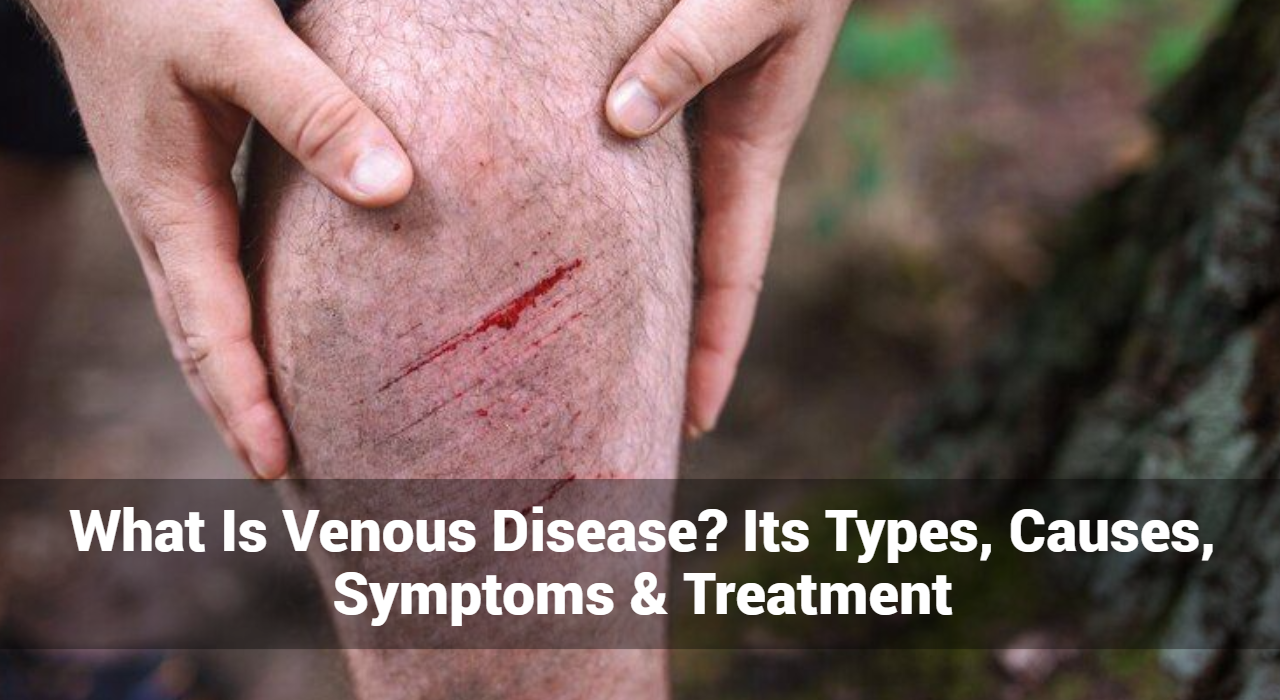

Different Types of Raynaud’s Disease: How to Treat Each One

Raynaud’s Disease, also known as Raynaud phenomenon or Raynaud’s syndrome, is a condition characterized by episodes of vasospasm, which causes reduced blood flow to certain areas of the body, typically the fingers and toes. These episodes can lead to color changes, numbness, and pain in the affected areas. Raynaud’s disease can be primary or secondary, each with distinct causes and treatment approaches. This article provides an in-depth look at the different types of Raynaud disease and how to treat each one effectively.

Different Types of Raynaud Disease

1. Primary Raynaud’s Disease

Primary Raynaud’s disease (also called Raynaud’s phenomenon) is the more common and less severe form. It occurs without any associated medical condition and is often less severe. It primarily affects young women and typically begins between the ages of 15 and 30.

Causes

  • Idiopathic: The exact cause is unknown.
  • Genetic Predisposition: There may be a hereditary component.

Symptoms

  • Color Changes: Fingers or toes turn white (blanching) or blue in response to cold or stress.
  • Numbness and Tingling: Reduced blood flow causes numbness and a prickly feeling.
  • Pain: Throbbing pain may occur as blood flow returns.

2. Secondary Raynaud’s Disease

Secondary Raynaud’s disease (also known as Raynaud’s syndrome) is less common but more severe. It is associated with other underlying conditions and usually affects older individuals.

Causes

  • Connective Tissue Diseases: Such as scleroderma, lupus, and rheumatoid arthritis.
  • Arterial Diseases: Atherosclerosis and Buerger’s disease.
  • Occupational Causes: Jobs involving vibrating tools or repetitive motions.
  • Medications: Some beta-blockers, chemotherapy agents, and migraine medications.
  • Injuries: Hand or foot injuries.

Symptoms

  • More Severe Episodes: Longer duration and more frequent episodes compared to primary Raynaud disease.
  • Ulcers and Infections: Due to prolonged lack of blood flow, ulcers or sores can develop.
  • Associated Symptoms: Symptoms of the underlying condition, such as joint pain or skin changes.

Track and Manage your Eczema treatment using a comprehensive Eczema App
Download Eczemaless now


General Treatment Approaches Management for Both Raynaud’s Disease Types

01. Lifestyle Modifications

  • Avoid Cold Exposure: Keep the entire body warm, especially hands and feet. Wear gloves, warm socks, and use hand warmers in cold weather.
  • Reduce Stress: Practice relaxation techniques such as yoga, meditation, or deep breathing exercises.
  • Avoid Smoking: Smoking can constrict blood vessels and worsen symptoms.

02. Diet and Supplements

  • Healthy Diet: A balanced diet rich in fruits, vegetables, whole grains, and lean proteins supports overall vascular health.
  • Omega-3 Fatty Acids: Found in fish oil, flaxseed oil, and walnuts, omega-3s can improve blood flow.
  • Ginkgo Biloba: This supplement may improve circulation.

What Are The Treatments for Primary Raynaud’s Disease?

01. Medications

  • Calcium Channel Blockers: Such as nifedipine and amlodipine, can help dilate blood vessels and reduce the frequency and severity of attacks.
  • Vasodilators: Drugs like nitroglycerin ointment can be applied to affected areas to widen blood vessels.

02. Behavioral Therapy

  • Biofeedback: This technique teaches control over body functions, such as blood flow, to help manage symptoms.
  • Cognitive Behavioral Therapy (CBT): Can help manage stress, which is a common trigger.

What Are The Treatments for Secondary Raynaud’s Disease?

01. Medications

  • Calcium Channel Blockers and Vasodilators: Similar to primary Raynaud disease, these can be effective.
  • ACE Inhibitors: Such as enalapril, can help reduce the frequency of attacks.
  • Prostaglandins: Intravenous prostaglandins like iloprost can help in severe cases by dilating blood vessels.

02. Treatment of Underlying Conditions

  • Connective Tissue Disease Management: Medications like immunosuppressants, corticosteroids, or antimalarial drugs to control the primary disease.
  • Addressing Arterial Conditions: Treatments such as angioplasty for atherosclerosis.

03. Surgical Options

  • Sympathectomy: A surgical procedure to cut the nerves that cause blood vessel constriction. This is considered when symptoms are severe and not responsive to other treatments.
  • Botox Injections: Botulinum toxin injections can block nerves that constrict blood vessels, relieving severe symptoms.

04. Advanced Therapies

  • Phosphodiesterase Inhibitors: Drugs like sildenafil (Viagra) can improve blood flow.
  • Endothelin Receptor Antagonists: Such as bosentan, can be used in severe cases, particularly when associated with scleroderma.


GET IN CONTROL OF YOUR ECZEMA

Use our AI tool to check the severity of Eczema and keep track of your Eczema progress.

Use our AI tool to check the severity of Eczema and keep track of your Eczema progress.


How to Help Prevent Raynaud’s Attacks?

Raynaud’s disease, characterized by episodes of reduced blood flow to the fingers and toes, can be both uncomfortable and disruptive. Prevention strategies focus on reducing triggers, enhancing blood flow, and maintaining overall vascular health. Here are effective methods to help prevent Raynaud’s attacks:

1. Lifestyle Modifications

Stay Warm

  • Dress Appropriately: Wear warm clothing, especially gloves, socks, and hats, in cold weather. Layer your clothing to trap body heat.
  • Use Warm Packs: Carry hand and foot warmers during cold weather or stressful situations.
  • Warm Environment: Keep your home and workplace warm. Use space heaters if necessary.

Manage Stress

  • Relaxation Techniques: Practice stress-reducing activities like yoga, meditation, or deep-breathing exercises.
  • Regular Exercise: Physical activity can improve circulation and reduce stress. Aim for at least 30 minutes of moderate exercise most days of the week.
  • Avoid Stressful Situations: Identify and manage situations that trigger stress. Consider professional counseling or stress management programs if needed.

2. Dietary Adjustments

Healthy Diet

  • Balanced Nutrition: Consume a diet rich in fruits, vegetables, whole grains, lean proteins, and healthy fats to support vascular health.
  • Omega-3 Fatty Acids: Include foods high in omega-3s, such as fatty fish (salmon, mackerel), flaxseeds, and walnuts. These can improve blood flow and reduce inflammation.

Hydration

  • Drink Plenty of Water: Staying hydrated is essential for maintaining good circulation. Aim for at least 8 glasses of water a day.

3. Avoiding Triggers

Cold Exposure

  • Limit Time Outside: Stay indoors during extreme cold weather.
  • Pre-warm Your Car: Start your car and let it warm up before driving in cold weather.
  • Warm Water: Wash hands and bathe with warm water instead of cold.

Vibrating Tools and Repetitive Movements

  • Protect Your Hands: Use protective gear if you must use vibrating tools or perform repetitive tasks.
  • Take Breaks: Regularly rest your hands during activities involving repetitive motions.

4. Smoking and Alcohol

Quit Smoking

  • Nicotine Effects: Nicotine constricts blood vessels and can trigger Raynaud’s attacks. Seek support to quit smoking through counseling, nicotine replacement therapies, or prescription medications.

Limit Alcohol Consumption

  • Moderation: Alcohol can affect blood circulation. Drink in moderation, if at all, and be aware of its impact on your symptoms.

5. Medications and Supplements

Consult Your Doctor

  • Review Medications: Some medications, such as beta-blockers and certain migraine drugs, can worsen Raynaud’s. Discuss alternatives with your healthcare provider.
  • Supplements: Consider supplements like ginkgo biloba or fish oil, which may improve circulation. Always consult with a healthcare professional before starting any new supplements.

6. Hands and Feet Care

Moisturize Regularly

  • Prevent Dryness: Use lotions and creams to keep your skin hydrated and prevent cracking, which can worsen symptoms.

Massage

  • Enhance Circulation: Gentle hand and foot massages can stimulate blood flow.

7. Regular Medical Checkups

Monitor Your Health

  • Regular Visits: Keep up with regular checkups to monitor your condition and adjust treatments as necessary.
  • Early Intervention: Address any new or worsening symptoms promptly with your healthcare provider.

Conclusion

Raynaud’s disease, whether primary or secondary, can significantly impact the quality of life. Understanding the differences between the two types is crucial for effective management. Primary Raynaud disease generally responds well to lifestyle modifications and medications, while secondary Raynaud disease often requires a more aggressive approach, including treatment of underlying conditions and possibly surgical interventions. If you suspect you have Raynaud disease, it is important to consult with a healthcare provider to receive a proper diagnosis and tailored treatment plan. With appropriate management, individuals with Raynaud’s disease can lead a comfortable and active life.


Track and Manage your Eczema treatment using a comprehensive Eczema App
Download Eczemaless now



Psoriasis on the Face: Causes, Symptoms, and Treatments

Psoriasis is a chronic skin condition that affects millions of people worldwide. While it can appear on various parts of the body, psoriasis on the face can be particularly challenging due to its visibility and the delicate nature of facial skin. Understanding the causes, recognizing the symptoms, and exploring effective treatments are crucial for managing this condition. This comprehensive guide delves into all aspects of facial psoriasis, providing valuable insights for those affected by this persistent skin issue.

What is Psoriasis?

Psoriasis is an autoimmune disorder characterized by the rapid turnover of skin cells, leading to the formation of thick, scaly patches. These patches, often referred to as plaques, can be itchy, red, and inflamed. While psoriasis can manifest anywhere on the body, facial psoriasis specifically impacts areas such as the eyebrows, upper forehead, hairline, and the skin between the nose and upper lip.

Causes of Psoriasis on the Face

The exact cause of psoriasis remains unknown, but it is believed to be a combination of genetic, environmental, and immune system factors. Here are some key factors contributing to psoriasis on the face:

  • Genetics: A family history of psoriasis increases the likelihood of developing the condition.
  • Immune System Dysfunction: Psoriasis is an autoimmune disorder where the immune system mistakenly attacks healthy skin cells, accelerating their production and leading to the formation of plaques.
  • Environmental Triggers: Factors such as stress, smoking, alcohol consumption, and certain medications can trigger or worsen psoriasis.
  • Infections: Infections, particularly streptococcal throat infections, can trigger facial psoriasis in some individuals.

Track and Manage your Eczema treatment using a comprehensive Eczema App
Download Eczemaless now
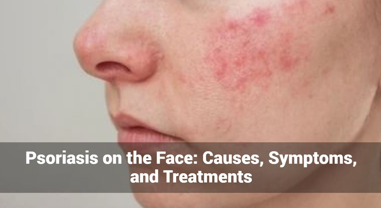

What Are The Symptoms of Psoriasis on the Face?

Identifying the symptoms of psoriasis on the face is crucial for early diagnosis and effective management. Common symptoms include:

  • Red Patches: Well-defined red patches on the facial skin.
  • Scales: Silvery-white scales covering the red patches.
  • Dry Skin: Excessive dryness leading to cracking and bleeding.
  • Itchiness and Burning: Persistent itching and a burning sensation on the affected areas.
  • Swelling: Inflammation and swelling around the plaques.

What Types of Psoriasis on the Face?

Psoriasis on the face can present in various forms, including:

  • Plaque Psoriasis: The most common type, characterized by raised, red patches with a silvery scale.
  • Guttate Psoriasis: Small, dot-like lesions, often triggered by infections.
  • Inverse Psoriasis: Smooth, red lesions that occur in skin folds.
  • Erythrodermic Psoriasis: A rare, severe form causing widespread redness and shedding of the skin.

Diagnosing Psoriasis on the Face

A dermatologist typically diagnoses psoriasis based on the appearance of the skin. In some cases, a skin biopsy may be performed to confirm the diagnosis and rule out other skin conditions. A thorough medical history and examination help identify potential triggers and co-existing conditions.

What Are The Treatment Options for Psoriasis on the Face?

Treating psoriasis on the face requires careful consideration due to the delicate nature of facial skin. The goal of treatment is to reduce inflammation, slow the rapid growth of skin cells, and clear plaques. Here are the main treatment options:

Topical Treatments

  1. Corticosteroids
    • Mild to Moderate Corticosteroids: These are often the first line of treatment. They reduce inflammation and slow down skin cell turnover. Examples include hydrocortisone and desonide.
    • Usage: Apply sparingly to affected areas, as long-term use can cause thinning of the skin and other side effects.
  2. Vitamin D Analogues
    • Calcipotriol (Calcipotriene) and Calcitriol: These help to normalize skin cell production and reduce scaling. They are less likely to cause skin thinning compared to corticosteroids.
    • Usage: Often used in combination with corticosteroids for enhanced effectiveness.
  3. Calcineurin Inhibitors
    • Tacrolimus (Protopic) and Pimecrolimus (Elidel): These are non-steroidal treatments that reduce inflammation and are suitable for sensitive areas like the face. They do not cause skin thinning.
    • Usage: Applied twice daily to affected areas, particularly useful for delicate skin regions.
  4. Moisturizers
    • Emollients and Hydrating Creams: Regular use of moisturizers helps to manage dryness, reduce scaling, and maintain skin hydration. Look for products free from fragrances and irritants.
    • Usage: Apply multiple times daily, especially after washing the face.


GET IN CONTROL OF YOUR ECZEMA

Use our AI tool to check the severity of Eczema and keep track of your Eczema progress.

Use our AI tool to check the severity of Eczema and keep track of your Eczema progress.

Phototherapy

  1. UVB Therapy
    • Narrowband UVB Therapy: Involves exposure to ultraviolet B light under medical supervision. It can slow down the growth of affected skin cells.
    • Usage: Typically performed in a dermatologist’s office, requiring multiple sessions per week.
  2. PUVA Therapy
    • Psoralen + UVA Therapy: Combines psoralen medication with exposure to UVA light. Psoralen makes the skin more sensitive to light, enhancing the effectiveness of UVA therapy.
    • Usage: Often reserved for more severe cases and requires careful monitoring due to potential side effects.

Systemic Treatments

For moderate to severe facial psoriasis, systemic treatments may be necessary. These are usually prescribed when topical treatments are insufficient.

  1. Oral Medications
    • Methotrexate: Reduces inflammation and slows skin cell production. Requires regular blood tests to monitor for side effects.
    • Cyclosporine: An immunosuppressant that can rapidly improve symptoms but is usually used short-term due to potential side effects.
    • Acitretin: A retinoid that normalizes skin cell growth. Not suitable for pregnant women due to risk of birth defects.
  2. Biologic Therapies
    • Adalimumab (Humira), Etanercept (Enbrel), Infliximab (Remicade): These target specific parts of the immune system to control inflammation and reduce symptoms.
    • Usage: Administered via injection or infusion, often requiring regular monitoring by a healthcare professional.

Lifestyle And Natural Home Remedies That Really Work Against Psoriasis

  1. Stress Management
    • Techniques: Yoga, meditation, and mindfulness can help manage stress, which is a known trigger for psoriasis flare-ups.
    • Impact: Reducing stress can lead to fewer flare-ups and improved overall well-being.
  2. Healthy Diet
    • Anti-inflammatory Foods: Include fruits, vegetables, whole grains, and fatty fish rich in omega-3 fatty acids. Avoid foods that can trigger inflammation, such as processed foods and sugary items.
    • Hydration: Drinking plenty of water helps maintain skin hydration and overall health.
  3. Skincare Routine
    • Gentle Cleansers: Use mild, fragrance-free cleansers to avoid irritation. Avoid hot water, as it can dry out the skin.
    • Regular Moisturizing: Apply moisturizers immediately after washing the face to lock in moisture. Use products specifically designed for sensitive skin.

Emerging Treatments

  1. Topical Janus Kinase (JAK) Inhibitors
    • Ruxolitinib Cream: An emerging treatment showing promise in reducing inflammation and skin cell production in psoriasis. Clinical trials are ongoing to establish its effectiveness and safety for facial psoriasis.
  2. Laser Therapy
    • Excimer Laser: Delivers targeted UVB light to affected areas, which can be effective for small patches of facial psoriasis. Requires multiple sessions.

Conclusion

Psoriasis on the face, while challenging, can be managed effectively with the right approach. Understanding the causes, recognizing the symptoms, and exploring various treatment options are essential steps in managing this condition. By adopting a holistic approach that includes medical treatments, lifestyle changes, and emotional support, individuals with facial psoriasis can lead a fulfilling life.

For more information and support, consult a dermatologist and consider joining psoriasis support communities. Managing psoriasis is a journey, and with the right resources, it is possible to navigate it successfully.


Track and Manage your Eczema treatment using a comprehensive Eczema App
Download Eczemaless now
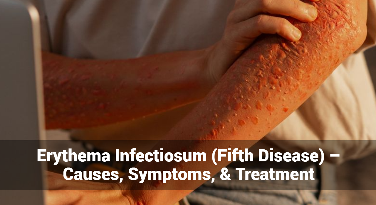


Erythema Infectiosum (Fifth Disease) – Causes, Symptoms, & Treatment

Erythema infectiosum, commonly known as fifth disease, is a highly contagious viral infection primarily affecting children. Recognized by its distinctive “slapped cheek” rash, erythema infectiosum is generally mild but can pose significant risks to certain populations. This article delves into the details of erythema infectiosum, exploring its causes, symptoms, and treatment.

What Is Erythema Infectiosum?

Erythema infectiosum is caused by the human parvovirus B19. It is one of several childhood exanthems (rashes) and is so named because it was historically the fifth disease in a classification of common childhood rashes. The condition is typically mild in healthy children but can have more severe implications for adults and individuals with weakened immune systems.

What Are The Causes of Erythema Infectiosum?

Erythema infectiosum, also known as fifth disease, is a viral infection primarily caused by the human parvovirus B19. This section will delve into the specific causes and contributing factors associated with erythema infectiosum.

Primary Cause: Human Parvovirus B19

Human parvovirus B19 is the sole causative agent of erythema infectiosum. This virus specifically targets and infects red blood cell precursors in the bone marrow, leading to a temporary disruption in red blood cell production. The infection process and subsequent immune response are what primarily lead to the symptoms observed in erythema infectiosum.

Transmission Methods

Erythema infectiosum spreads primarily through the following means:

  1. Respiratory Droplets: The most common transmission route is via respiratory droplets. When an infected person coughs or sneezes, they release tiny droplets containing the virus into the air. These droplets can be inhaled by nearby individuals, leading to infection.
  2. Direct Contact: The virus can also spread through direct contact with respiratory secretions. For example, sharing utensils, drinks, or touching surfaces contaminated with these secretions can facilitate the spread of the virus.
  3. Blood Transmission: Less commonly, human parvovirus B19 can be transmitted through blood or blood products. This can occur through transfusions, organ transplants, or shared needles.
  4. Vertical Transmission: Pregnant women infected with human parvovirus B19 can transmit the virus to their fetus. This vertical transmission can lead to severe complications such as fetal anemia or hydrops fetalis.

Track and Manage your Eczema treatment using a comprehensive Eczema App
Download Eczemaless now

Factors Contributing to the Spread

Several factors contribute to the spread and impact of erythema infectiosum:

  1. Seasonal Variation: Outbreaks of erythema infectiosum are more common in late winter and early spring, although the virus can circulate year-round.
  2. Close Contact Environments: Settings such as schools, daycare centers, and households where individuals are in close proximity to each other facilitate the spread of the virus. Children are particularly susceptible due to their close interactions and developing immune systems.
  3. Immunocompromised Individuals: People with weakened immune systems, including those undergoing chemotherapy, organ transplant recipients, or individuals with HIV, are more susceptible to infection and may experience more severe symptoms.
  4. Chronic Anemia Conditions: Individuals with chronic anemia conditions, such as sickle cell disease or hereditary spherocytosis, are at higher risk for complications if they contract erythema infectiosum. The virus can exacerbate their condition by further disrupting red blood cell production.

Pathogenesis

The pathogenesis of erythema infectiosum involves several stages:

  1. Viral Entry and Replication: The human parvovirus B19 enters the body through the respiratory tract and initially replicates in the nasopharynx.
  2. Viremia: The virus then enters the bloodstream, leading to viremia. During this stage, the infected individual may experience flu-like symptoms.
  3. Immune Response: The body’s immune response to the virus results in the characteristic symptoms of erythema infectiosum, including the distinctive rash and joint pain. The immune response also helps to clear the virus from the body.
  4. Bone Marrow Suppression: Human parvovirus B19 targets red blood cell precursors in the bone marrow, temporarily halting their production. This effect is usually mild and transient in healthy individuals but can be severe in those with pre-existing anemia conditions.

Erythema infectiosum is caused by the human parvovirus B19, which spreads primarily through respiratory droplets, direct contact with infected secretions, blood transmission, and vertical transmission from mother to fetus.

What Are The Symptoms of Erythema Infectiosum?

Erythema infectiosum, commonly known as fifth disease, is a viral infection with a distinctive set of symptoms that evolve through different stages of the illness. Understanding these symptoms helps in early recognition and appropriate management of the condition.

Incubation Period

The incubation period for erythema infectiosum ranges from 4 to 14 days, although it can extend to 21 days in some cases. During this time, the infected person may not exhibit any noticeable symptoms.

Early Symptoms

Initial symptoms are often mild and nonspecific, resembling those of a common cold or flu. These can include:

  • Mild Fever: A low-grade fever is common, typically around 101°F (38.3°C).
  • Headache: Generalized headache.
  • Flu-like Symptoms: Including fatigue, muscle aches, and malaise.
  • Sore Throat: Mild throat irritation or soreness.
  • Runny or Stuffy Nose: Symptoms similar to a mild upper respiratory infection.

Rash Development

The hallmark of erythema infectiosum is its characteristic rash, which progresses through distinct stages:

  1. Slapped Cheek” Rash:
    • Appearance: Bright red rash on both cheeks, giving the appearance of slapped cheeks.
    • Timing: This facial rash typically appears after the early symptoms have subsided and the fever has resolved.
    • Common in Children: This symptom is most prominent in children and less common in adults.
  2. Body Rash:
    • Appearance: A lacy, red rash that can spread from the face to the trunk, arms, and legs.
    • Pattern: The rash has a reticular (net-like) pattern, often described as lacy or “fishnet-like.”
    • Itching: The body rash may be itchy, though the severity of itching varies.
  3. Reappearance:
    • Triggers: The rash can fade and reappear over several weeks, often triggered by factors such as sunlight exposure, heat, exercise, or stress.
    • Fluctuation: The intensity and distribution of the rash can fluctuate, appearing more pronounced under certain conditions.

Joint Pain and Swelling

Joint involvement is more common in adults than in children and can include:

  • Arthralgia: Pain in the joints, commonly affecting the hands, wrists, knees, and ankles.
  • Arthritis: In some cases, there may be swelling and inflammation of the joints, leading to arthritis-like symptoms.
  • Duration: Joint pain and swelling can last from a few days to several weeks or even months, but they typically resolve without long-term damage.

Additional Symptoms

In addition to the classic rash and joint symptoms, erythema infectiosum can present with other signs, especially in adults or those with compromised immune systems:

  • Generalized Fatigue: Persistent tiredness and lack of energy.
  • Gastrointestinal Symptoms: Mild nausea or abdominal discomfort in some cases.
  • Respiratory Symptoms: Cough and mild respiratory distress.


GET IN CONTROL OF YOUR ECZEMA

Use our AI tool to check the severity of Eczema and keep track of your Eczema progress.

Use our AI tool to check the severity of Eczema and keep track of your Eczema progress.


Treatment of Erythema Infectiosum

While it usually resolves on its own without specific antiviral treatment, managing the symptoms can provide relief and prevent complications. Here’s an in-depth look at the treatment strategies for erythema infectiosum.

General Management

The cornerstone of treating erythema infectiosum involves supportive care to alleviate symptoms and ensure comfort. Here are the key approaches:

Symptomatic Relief

  • Fever and Pain: Over-the-counter medications such as acetaminophen (Tylenol) or ibuprofen (Advil, Motrin) can be used to reduce fever and relieve pain, including headache and joint discomfort.
  • Itching: Antihistamines (such as diphenhydramine or cetirizine) may help reduce itching associated with the rash.

Hydration and Rest

  • Fluids: Encourage adequate fluid intake to stay hydrated, especially if the patient has a fever.
  • Rest: Ensure plenty of rest to help the body recover from the viral infection.

Skin Care

  • Moisturizers: Use gentle moisturizers to soothe irritated skin.
  • Avoid Triggers: Minimize exposure to sunlight, heat, and activities that might exacerbate the rash.

Specific Considerations for Different Populations

Children

  • Comfort Measures: Ensuring comfort through appropriate clothing and a cool environment can help manage symptoms.
  • Monitoring: Keep an eye on symptoms and ensure they don’t escalate, particularly if the child has underlying health issues.

Adults

  • Joint Pain Management: Since joint pain and swelling are more common in adults, NSAIDs like ibuprofen can be particularly helpful. In some cases, more potent anti-inflammatory medications may be needed.

Pregnant Women

  • Monitoring: Pregnant women who contract erythema infectiosum should be closely monitored due to the risk of fetal complications. Regular ultrasounds may be recommended to check for signs of fetal distress, such as hydrops fetalis.
  • Medical Consultation: Immediate consultation with a healthcare provider is essential if a pregnant woman is exposed to or develops symptoms of erythema infectiosum.

Immunocompromised Individuals

  • Medical Management: Those with weakened immune systems, such as patients undergoing chemotherapy or those with HIV, may require more intensive medical care. This can include antiviral medications and treatments to support the immune system.
  • Monitoring for Complications: Close monitoring for potential complications, such as severe anemia, is crucial.

Individuals with Chronic Anemia

  • Aplastic Crisis Management: Individuals with health conditions like sickle cell disease or other hemolytic anemias are at risk for aplastic crises. Treatment may include blood transfusions and hospitalization to manage severe anemia.
  • Regular Check-ups: Ongoing monitoring and supportive care to manage their underlying condition and prevent complications.

Preventive Measures

Good Hygiene Practices

  • Hand Washing: Regular and thorough hand washing with soap and water can help prevent the spread of the virus.
  • Respiratory Etiquette: Covering the mouth and nose with a tissue or elbow when coughing or sneezing can reduce the risk of transmission.

Avoiding Exposure

  • Stay Home: Infected individuals, especially children, should stay home from school or daycare during the contagious period to prevent spreading the virus to others.
  • Isolate: Avoid close contact with others, particularly vulnerable groups such as pregnant women and immunocompromised individuals.

When to Seek Medical Attention

While erythema infectiosum is typically mild, certain situations warrant medical attention:

  • Severe Symptoms: Persistent high fever, severe headache, or intense joint pain.
  • Complications: Signs of complications such as difficulty breathing, significant swelling, or prolonged symptoms.
  • Pregnancy: If a pregnant woman is exposed to the virus or develops symptoms.
  • Immunocompromised Individuals: Any signs of severe illness or complications in individuals with weakened immune systems.

Prevention of Erythema Infectiosum

Preventive measures focus on good hygiene and avoiding exposure to the virus:

  • Good Hygiene Practices: Frequent handwashing, avoiding close contact with infected individuals, and proper respiratory etiquette (covering mouth and nose when coughing or sneezing).
  • Avoiding Exposure: Pregnant women and immunocompromised individuals should avoid contact with known cases of erythema infectiosum.

Conclusion

Erythema infectiosum is a common viral infection marked by its characteristic rash and mild flu-like symptoms. While typically mild, it can lead to complications in pregnant women, immunocompromised individuals, and those with chronic anemia. Understanding the causes, symptoms, and treatment options for erythema infectiosum can help manage and prevent the spread of this infection effectively. With proper care and preventive measures, the impact of erythema infectiosum can be minimized, ensuring better health outcomes for affected individuals.


Track and Manage your Eczema treatment using a comprehensive Eczema App
Download Eczemaless now

What Is Inverse Psoriasis: It’s Symptoms, Causes, & Effective Treatments

Inverse psoriasis is a lesser-known form of psoriasis that manifests in skin folds and creases, often presenting unique challenges in diagnosis and management. In this comprehensive guide, we’ll delve into the symptoms, causes, and treatment options for inverse psoriasis, offering valuable insights for individuals grappling with this condition.

What is Inverse Psoriasis?

Inverse psoriasis, also known as intertriginous psoriasis, is a subtype of psoriasis characterized by smooth, red, inflamed patches of skin that occur in the skin folds and creases of the body. Unlike other forms of psoriasis, such as plaque psoriasis which typically appears on the elbows, knees, and scalp, inverse psoriasis tends to affect areas like the armpits, groin, under the breasts, and around the genitals.

How Do Symptoms Of Inverse Psoriasis Impact Daily Activities And Quality Of Life?

Symptoms of inverse psoriasis often manifest as:

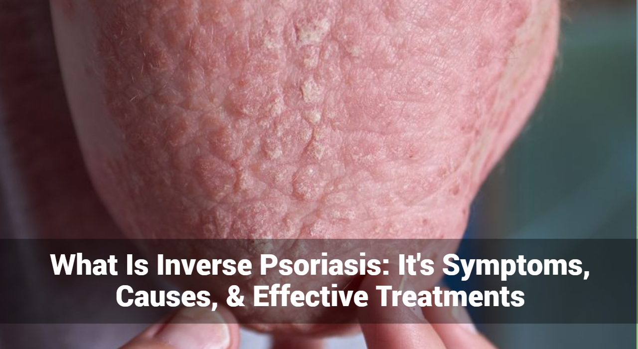
  1. Smooth, Red Patches: Inverse psoriasis typically presents as smooth, red patches of inflamed skin. Unlike the raised, scaly patches commonly seen in other forms of psoriasis, the lesions in inverse psoriasis are often flat and may have a shiny appearance.
  2. Skin Folds and Creases: Affected areas tend to occur in skin folds and creases, such as the armpits, groin, under the breasts, and around the genitals. These regions provide an ideal environment for the development of inverse psoriasis due to increased friction and moisture.
  3. Itching and Discomfort: Individuals with inverse psoriasis may experience itching, burning, or soreness in the affected areas. Discomfort can be exacerbated by friction from clothing or physical activity.
  4. Moist Appearance: Due to the moisture buildup in skin folds, areas affected by inverse psoriasis may appear moist or shiny. This moisture can further contribute to skin irritation and discomfort.
  5. Susceptibility to Fungal Infections: The warm, moist environment of skin folds makes them susceptible to fungal infections. Individuals with inverse psoriasis may be more prone to developing secondary fungal infections, such as candidiasis, in affected areas.
  6. Aggravation by Sweating: Sweat can exacerbate symptoms of inverse psoriasis, leading to increased itching and discomfort. Managing sweating through proper hygiene and avoiding tight clothing can help alleviate symptoms.

It’s important to note that symptoms of inverse psoriasis can vary from person to person and may fluctuate in severity over time. If you suspect you may have inverse psoriasis or are experiencing persistent skin symptoms, it’s advisable to consult a dermatologist or healthcare professional for accurate diagnosis and personalized treatment recommendations.


Track and Manage your Eczema treatment using a comprehensive Eczema App
Download Eczemaless now


What Are The Causes Of Inverse Psoriasis?

Inverse psoriasis, like other forms of psoriasis, is a complex skin conditions with multiple factors contributing to its onset. Here’s an exploration of the causes of inverse psoriasis:

  1. Genetic Predisposition: Genetic factors play a significant role in predisposing individuals to inverse psoriasis. Research suggests that certain genetic variations increase susceptibility to psoriasis, including inverse psoriasis.
  2. Immune System Dysfunction: Inverse psoriasis is considered an autoimmune disorder, where the immune system mistakenly targets healthy skin cells, leading to inflammation and accelerated skin cell growth. This immune system dysfunction is a key driver of psoriasis, including its inverse form.
  3. Environmental Triggers: External factors such as stress, infections (e.g., streptococcal infections), injury to the skin, or certain medications can trigger flare-ups or worsen existing inverse psoriasis. Environmental triggers vary among individuals and can play a significant role in the onset and severity of the condition.
  4. Obesity and Skin Folds: Obesity is associated with an increased risk of developing psoriasis, including inverse psoriasis. Excess weight can lead to friction and sweating in skin folds, creating an ideal environment for psoriatic lesions to develop and worsen. The interaction between obesity and psoriasis is complex and may involve inflammation and metabolic factors.
  5. Hormonal Factors: Hormonal changes, such as those occurring during puberty, pregnancy, or menopause, can influence the onset or severity of psoriasis, including inverse psoriasis. Fluctuations in hormone levels may affect immune function and inflammatory responses, potentially exacerbating psoriatic symptoms.
  6. Lifestyle Factors: Factors such as smoking, excessive alcohol consumption, poor diet, and sedentary behavior can contribute to inflammation and immune dysregulation, potentially worsening psoriasis symptoms, including those of the inverse form.
  7. Psychological Factors: Stress, anxiety, and depression can exacerbate psoriasis symptoms through the release of stress hormones and modulation of immune responses. Managing stress and maintaining good mental health can be crucial in managing inverse psoriasis effectively.

Understanding these various causes of inverse psoriasis can help individuals and healthcare professionals develop comprehensive management strategies tailored to each individual’s unique needs.

Diagnosis of Inverse Psoriasis

Diagnosing inverse psoriasis typically involves a thorough examination of the affected skin by a dermatologist or healthcare professional. Since inverse psoriasis can resemble other skin conditions such as fungal infections or intertrigo, a skin biopsy may be performed to confirm the diagnosis and rule out other potential causes.

GET IN CONTROL OF YOUR ECZEMA

Use our AI tool to check the severity of Eczema and keep track of your Eczema progress.

Use our AI tool to check the severity of Eczema and keep track of your Eczema progress.

What Are The Treatment Options for Inverse Psoriasis?

Treatment for inverse psoriasis aims to reduce inflammation, alleviate symptoms, and prevent flare-ups. Common treatment options include:

Topical Treatments:

  • Corticosteroids: Topical corticosteroids are commonly prescribed to reduce inflammation and itching in affected areas. They come in varying strengths and formulations, depending on the severity of the symptoms.
  • Calcineurin Inhibitors: Tacrolimus and pimecrolimus are calcineurin inhibitors that can be applied topically to reduce inflammation and help manage inverse psoriasis symptoms, particularly in sensitive areas.
  • Coal Tar Preparations: Coal tar products can help reduce inflammation and scaling associated with psoriasis. They are available in various formulations, including creams, ointments, and shampoos.

Phototherapy:

  • UVB Phototherapy: Exposure to ultraviolet B (UVB) light can help slow the growth of skin cells and reduce inflammation in inverse psoriasis lesions. UVB phototherapy may be administered in a dermatologist’s office or through at-home phototherapy devices under medical supervision.
  • PUVA Therapy: Psoralen plus ultraviolet A (PUVA) therapy involves taking a light-sensitizing medication (psoralen) before exposure to ultraviolet A (UVA) light. PUVA therapy can be effective in treating inverse psoriasis, but it requires careful monitoring due to potential side effects.

Systemic Medications:

  • Oral Retinoids: Oral retinoids, such as acitretin, can help reduce inflammation and slow down the growth of skin cells in inverse psoriasis. They are typically reserved for severe cases due to their potential side effects.
  • Methotrexate: Methotrexate is an immunosuppressive medication that can help control inflammation and reduce psoriasis symptoms, including those of inverse psoriasis.
  • Cyclosporine: Cyclosporine is another immunosuppressive medication that may be prescribed for severe cases of inverse psoriasis. It works by suppressing the immune system’s response that leads to inflammation.

Biologic Therapies:

  • TNF-alpha Inhibitors: Biologic drugs, such as adalimumab, etanercept, and infliximab, target specific molecules in the immune system involved in the inflammatory process of psoriasis. They are administered via injection or infusion and can be highly effective in treating inverse psoriasis.
  • IL-17 Inhibitors: Drugs that target interleukin-17 (IL-17), such as secukinumab and ixekizumab, have shown efficacy in treating inverse psoriasis by reducing inflammation and improving symptoms.

Lifestyle and Home Remedies For Inverse Psoriasis

In addition to medical treatments, certain lifestyle modifications and home remedies may help manage inverse psoriasis symptoms, including:

  • Maintaining Skin Hygiene: Keeping the affected areas clean and dry can help prevent bacterial and fungal infections, which can exacerbate inverse psoriasis.
  • Avoiding Irritants: Avoiding harsh soaps, perfumes, and other potential irritants can help minimize skin irritation and inflammation.
  • Moisturizing: Regularly moisturizing the skin can help alleviate dryness and reduce itching associated with inverse psoriasis.
  • Stress Management: Managing stress through relaxation techniques, meditation, or therapy can help reduce flare-ups of inverse psoriasis, as stress is known to exacerbate symptoms.

Conclusion:

Inverse psoriasis can pose significant challenges for individuals due to its location in skin folds and creases, but with proper diagnosis and treatment, symptoms can be effectively managed. By understanding the symptoms, causes, and treatment options for inverse psoriasis, individuals can take proactive steps towards achieving healthier skin and improved quality of life.

In conclusion, inverse psoriasis is a chronic skin condition that requires ongoing management, but with the right approach, individuals can find relief from symptoms and lead fulfilling lives. If you suspect you may have inverse psoriasis or are experiencing persistent skin symptoms, consult a dermatologist or healthcare professional for personalized evaluation and treatment recommendations.


Track and Manage your Eczema treatment using a comprehensive Eczema App
Download Eczemaless now


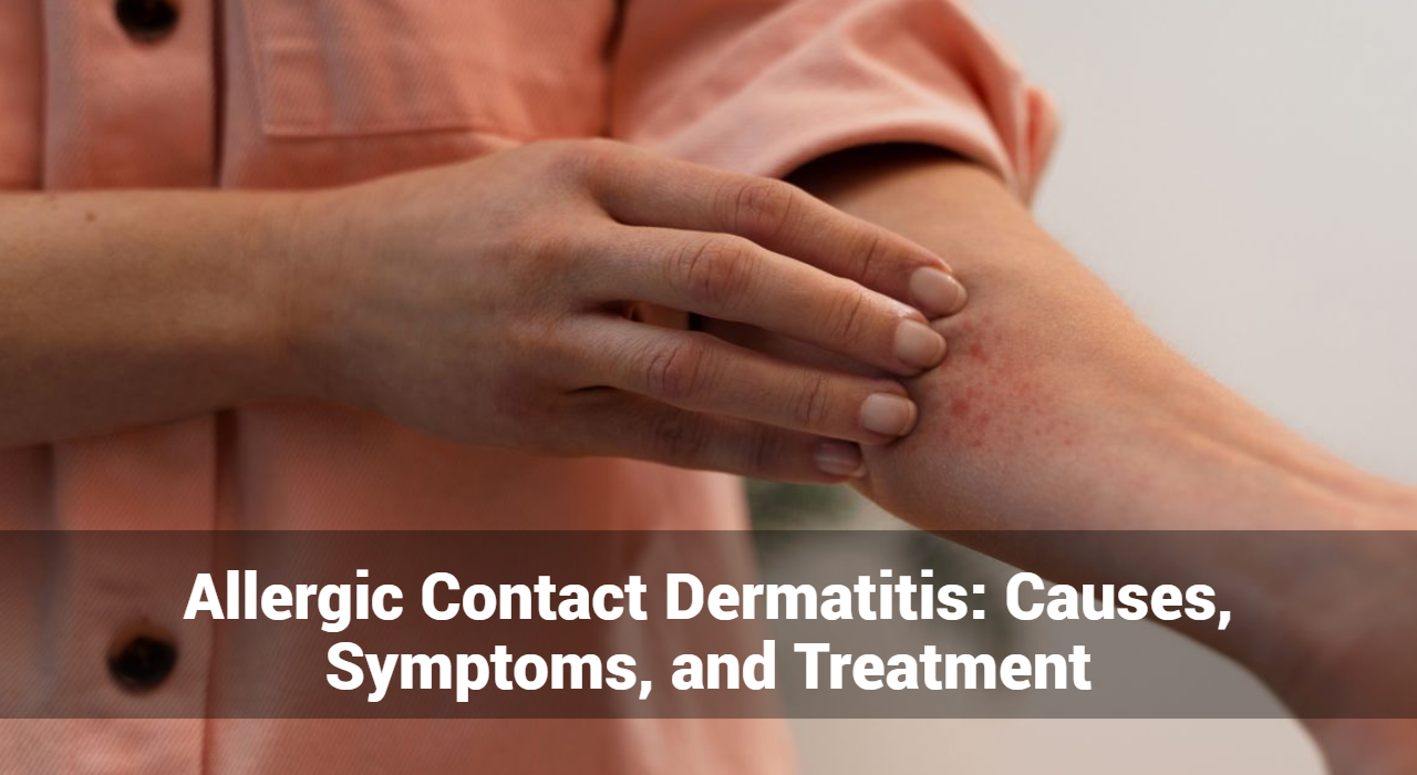
Allergic Contact Dermatitis: Causes, Symptoms, and Treatment

Allergic Contact Dermatitis (ACD) is a prevalent skin condition that significantly impacts quality of life. It arises from an allergic reaction when the skin comes into contact with specific substances, known as allergens. Understanding the causes, symptoms, and treatments of ACD is crucial for effective management and prevention. This article provides an in-depth look at ACD, focusing on its causes, clinical manifestations, diagnostic methods, and treatment options.

What is Allergic Contact Dermatitis?

Allergic Contact Dermatitis is an inflammatory skin condition caused by an allergic reaction to a substance in contact with the skin. Unlike irritant contact dermatitis, which occurs due to direct chemical damage to the skin, ACD is an immune-mediated response. This means that the body’s immune system reacts against a specific substance, leading to skin inflammation.

What Causes of Allergic Contact Dermatitis?

Common Allergens

01. Metals

  • Nickel: Found in jewelry, belt buckles, and eyeglass frames, nickel is one of the most common causes of ACD.
  • Cobalt: Often used in metal alloys and found in certain dyes and pigments.
  • Chromium: Present in cement, leather products, and some paints.

02. Fragrances

  • Used in perfumes, cosmetics, soaps, and detergents. Fragrances are a common cause of ACD, and many people are sensitive to them.

03. Preservatives

  • Formaldehyde: Used in cosmetics, disinfectants, and household cleaning products.
  • Methylisothiazolinone: Found in various personal care products and industrial applications.

04. Rubber Chemicals

  • Chemicals used in the production of rubber gloves, footwear, and other rubber products can cause ACD.

05. Plant Extracts

  • Poison Ivy, Oak, and Sumac: Contact with these plants can cause a severe allergic reaction in sensitive individuals.

06. Topical Medications

  • Neomycin: An antibiotic found in many over-the-counter creams.
  • Benzocaine: A local anesthetic used in various topical products.

Occupational Allergens

Certain professions are at higher risk of developing ACD due to frequent exposure to specific allergens:

01. Hairdressers and Cosmetologists

  • Exposed to hair dyes, bleaches, and preservatives.

02. Healthcare Workers

  • Regular use of latex gloves and disinfectants.

03. Construction Workers

  • Contact with cement, epoxy resins, and other building materials.

04. Agricultural Workers

  • Exposure to pesticides, fertilizers, and plant allergens.

Environmental and Lifestyle Factors

01. Climate and Pollution

  • Environmental factors like humidity and air pollution can exacerbate ACD symptoms.

02. Personal Care Products

  • Frequent use of fragranced and preservative-laden products can increase the risk of ACD.

Track and Manage your Eczema treatment using a comprehensive Eczema App
Download Eczemaless now


Symptoms of Allergic Contact Dermatitis

Acute Symptoms

  • Redness (Erythema): The affected area of the skin often becomes red and inflamed. This redness is a result of increased blood flow to the area as the body responds to the allergen.
  • Itching (Pruritus): Intense itching is one of the hallmark symptoms of ACD. This itching can be severe and persistent, leading to significant discomfort.
  • Swelling (Edema): The skin may swell, especially at the site of allergen contact. This swelling is due to the inflammatory response triggered by the immune system.
  • Blisters and Vesicles: Small fluid-filled blisters or vesicles can form on the skin. These blisters may burst, releasing their contents and potentially leading to crusting and oozing.
  • Pain and Tenderness: The affected area may become painful or tender to the touch. This symptom often accompanies severe cases of ACD.
  • Burning Sensation: Some individuals experience a burning or stinging sensation on the affected skin. This symptom can be particularly distressing and may indicate a more intense reaction.

Chronic Symptoms

  • Dry, Cracked Skin: Prolonged exposure to allergens or repeated episodes of ACD can cause the skin to become dry and cracked. This condition, known as xerosis, often leads to discomfort and an increased risk of secondary infections.
  • Thickened Skin (Lichenification): Chronic scratching and rubbing of the affected area can cause the skin to thicken and develop a leathery texture. This condition is known as lichenification and is often seen in long-standing cases of ACD.
  • Scaling and Flaking: The skin may start to scale and flake, shedding dry, dead skin cells. This symptom is common in chronic ACD and can be particularly noticeable on the hands and feet.
  • Hyperpigmentation or Hypopigmentation: Changes in skin color can occur, with the affected area becoming either darker (hyperpigmentation) or lighter (hypopigmentation) than the surrounding skin. These changes are often more apparent in individuals with darker skin tones.

Affected Areas

  • Hands: ACD commonly affects the hands, especially in individuals exposed to allergens through occupational or daily activities. Symptoms on the hands can significantly impact daily functioning.
  • Face and Neck: Allergens from cosmetics, fragrances, and jewelry often affect the face and neck. Symptoms in these areas can be particularly distressing due to their visibility.
  • Eyelids: The delicate skin of the eyelids is prone to ACD, especially from allergens in eye makeup, facial cleansers, or airborne substances.
  • Feet: Footwear materials, such as rubber or leather, can cause ACD on the feet. Symptoms may include itching, redness, and blistering on the soles and sides of the feet.
  • Other Common Sites: Other common sites include the wrists, due to watches and bracelets; the ears, due to earrings; and the torso, from clothing and belts.


GET IN CONTROL OF YOUR ECZEMA

Use our AI tool to check the severity of Eczema and keep track of your Eczema progress.

Use our AI tool to check the severity of Eczema and keep track of your Eczema progress.

Diagnosis of Allergic Contact Dermatitis

Clinical Evaluation

  1. Patient History: A detailed history of the patient’s exposure to potential allergens is essential for diagnosis.
  2. Physical Examination: Examining the characteristics and distribution of the rash helps in identifying ACD.

Patch Testing

  1. Procedure: Small amounts of suspected allergens are applied to the skin under occlusion and left for 48 hours.
  2. Interpretation: The test sites are examined for signs of an allergic reaction, such as redness, swelling, and vesiculation.

Differential Diagnosis

  • Irritant Contact Dermatitis: Caused by direct chemical damage to the skin, not an immune response.
  • Atopic Dermatitis: A chronic, genetic condition often accompanied by a history of allergies or asthma.
  • Other Skin Conditions: Conditions like psoriasis, seborrheic dermatitis, and fungal infections need to be ruled out.

Treatment of Allergic Contact Dermatitis

Avoidance of Allergens

01. Identification

  • Identifying and avoiding the specific allergen is the most effective way to manage ACD.

02. Protective Measures

  • Using protective clothing, gloves, and barrier creams can help minimize exposure to allergens.

Pharmacological Treatments

01. Topical Corticosteroids

  • Used to reduce inflammation and alleviate itching.

02. Topical Calcineurin Inhibitors

  • An alternative to corticosteroids for reducing inflammation.

03. Systemic Treatments

  • Oral corticosteroids or antihistamines may be prescribed for severe cases.

Non-Pharmacological Treatments

01. Phototherapy

  • Ultraviolet light therapy can help reduce inflammation and alleviate symptoms.

02. Alternative Therapies

  • Approaches like acupuncture, herbal treatments, and dietary changes may provide relief for some patients.

Management of Chronic ACD

01. Long-term Treatment Strategies

  • Continuous avoidance of allergens and regular use of emollients to maintain skin barrier function.

02. Patient Education

  • Educating patients about their condition and how to avoid triggers is crucial for effective management.

Prevention of Allergic Contact Dermatitis

Personal Care

01. Choosing Hypoallergenic Products

  • Using products labeled as hypoallergenic or fragrance-free can reduce the risk of ACD.

02. Patch Testing New Products

  • Testing new skincare or cosmetic products on a small skin area before full application.

Occupational Safety

01. Workplace Policies

  • Implementing safety measures to reduce exposure to allergens in the workplace.

02. Protective Equipment

  • Using gloves, masks, and protective clothing to minimize skin contact with allergens.

Public Awareness

01. Education Campaigns

  • Raising awareness about ACD and its causes through public health campaigns.

02. Support Groups

  • Providing support and resources for individuals affected by ACD.

Conclusion

Allergic Contact Dermatitis is a complex condition with significant impacts on individuals’ quality of life. Understanding its causes, symptoms, and treatment options is crucial for effective management and prevention. Through continued research, public awareness, and education, the burden of ACD can be reduced, leading to better outcomes for affected individuals. By identifying and avoiding allergens, utilizing appropriate treatments, and implementing preventive measures, those with ACD can manage their condition and improve their quality of life.


Track and Manage your Eczema treatment using a comprehensive Eczema App
Download Eczemaless now